Federal Institute of Piauí, Teresina 64000-040, Brazil.
Institute of Computing, Fluminense Federal University, Niterói, Rio de Janeiro 24220-900, Brazil.
Sensors (Basel). 2020 Jul 10;20(14):3866. doi: 10.3390/s20143866.
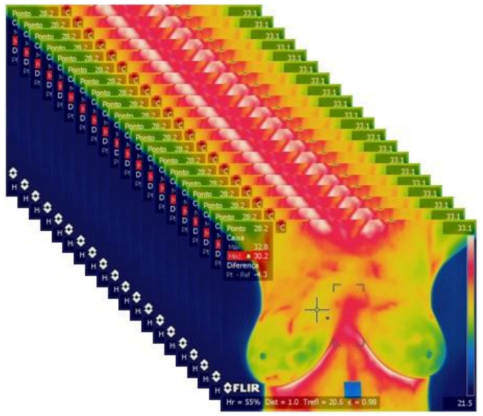
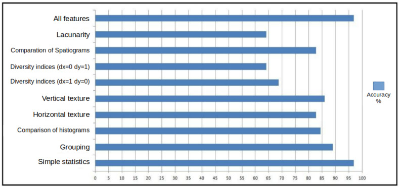
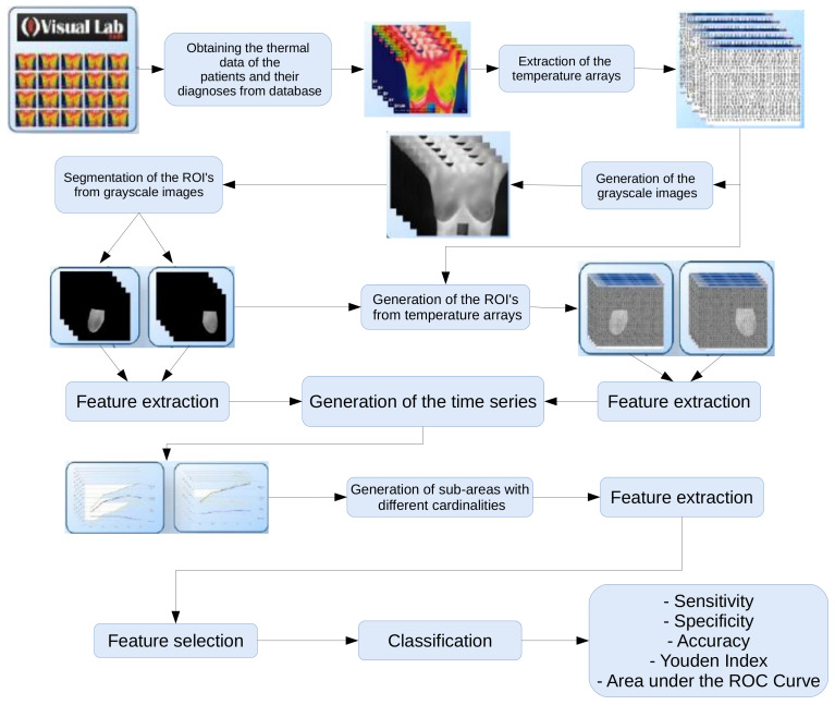
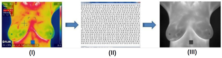
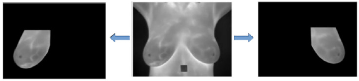
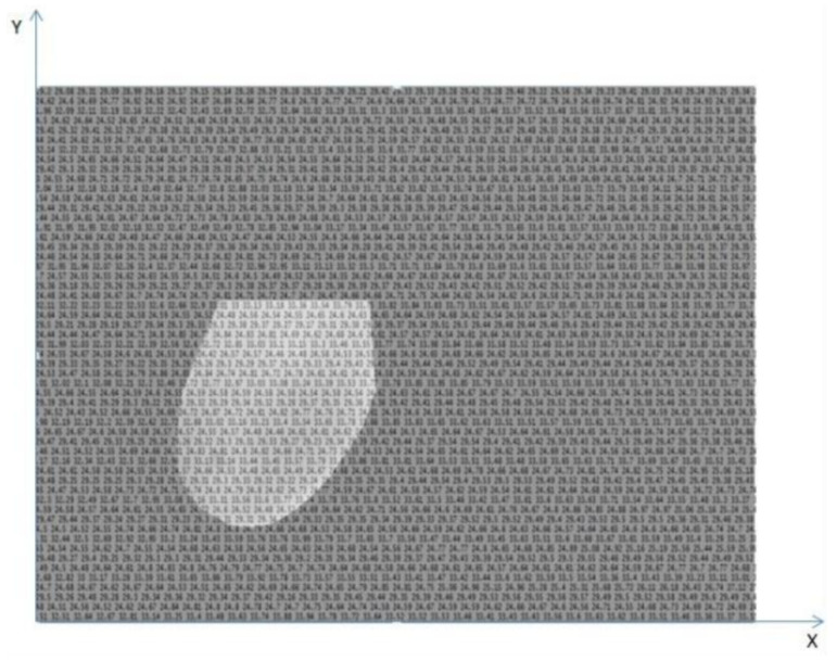
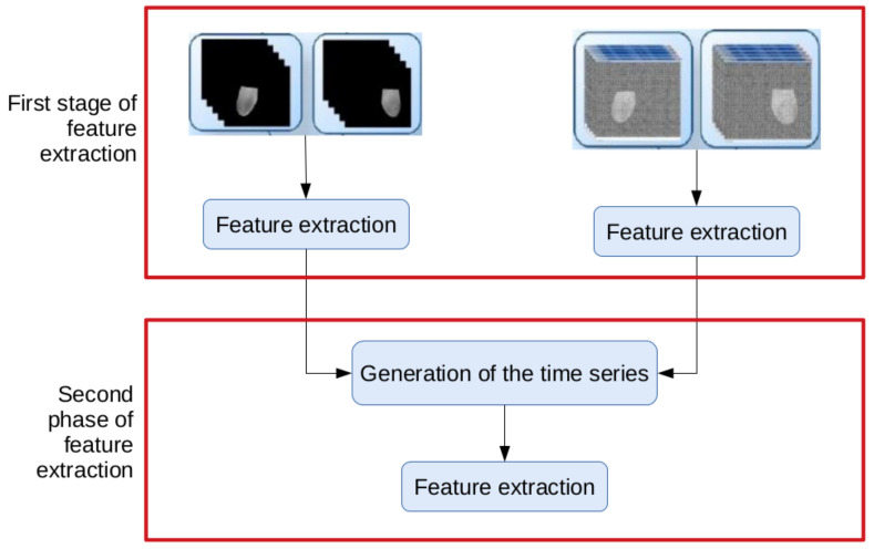
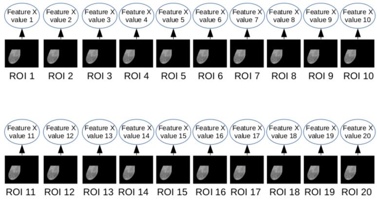
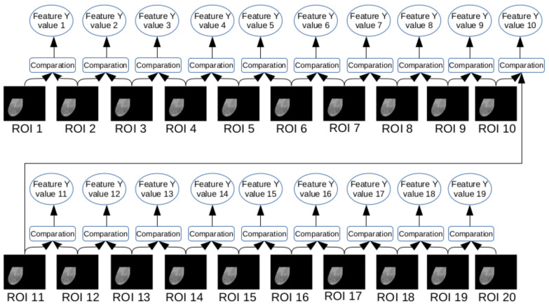
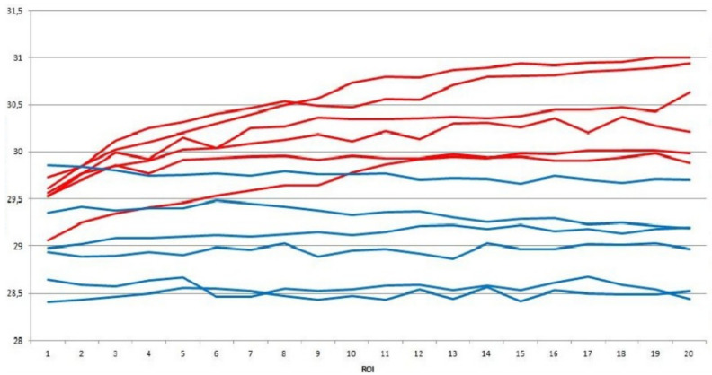
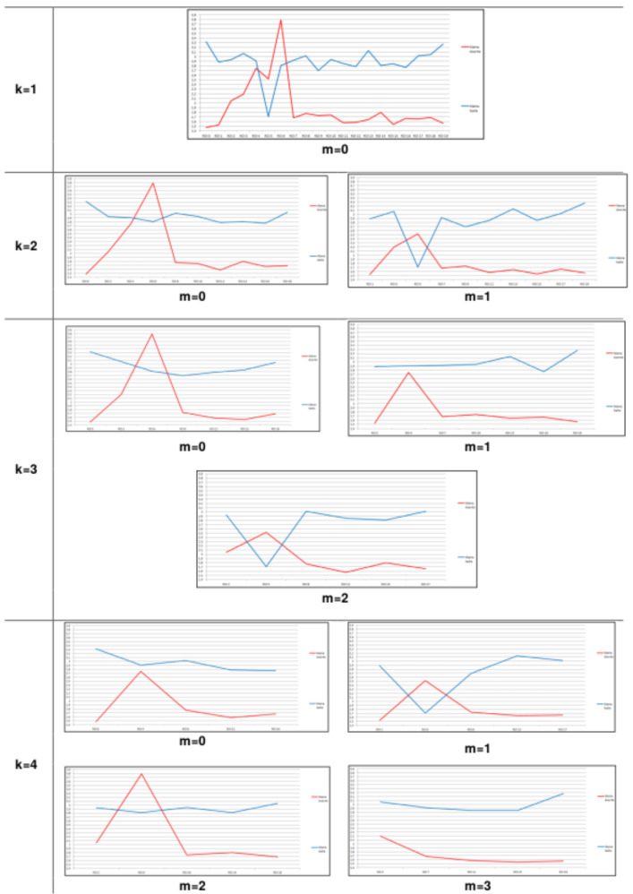
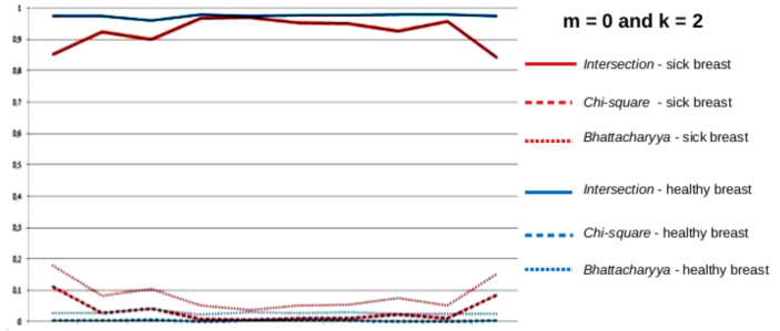
Breast cancer has been the second leading cause of cancer death among women. New techniques to enhance early diagnosis are very important to improve cure rates. This paper proposes and evaluates an image analysis method to automatically detect patients with breast benign and malignant changes (tumors). Such method explores the difference of Dynamic Infrared Thermography (DIT) patterns observed in patients' skin. After obtaining the sequential DIT images of each patient, their temperature arrays are computed and new images in gray scale are generated. Then the regions of interest (ROIs) of those images are segmented and, from them, arrays of the ROI temperature are computed. Features are extracted from the arrays, such as the ones based on statistical, clustering, histogram comparison, fractal geometry, diversity indices and spatial statistics. Time series that are broken down into subsets of different cardinalities are generated from such features. Automatic feature selection methods are applied and used in the Support Vector Machine (SVM) classifier. In our tests, using a dataset of 68 images, 100% accuracy was achieved.
乳腺癌是女性癌症死亡的第二大主要原因。提高早期诊断的新技术对于提高治愈率非常重要。本文提出并评估了一种图像分析方法,以自动检测患有乳腺良性和恶性变化(肿瘤)的患者。该方法探索了在患者皮肤中观察到的动态红外热成像(DIT)模式的差异。在获得每个患者的连续 DIT 图像后,计算其温度数组并生成灰度新图像。然后对这些图像的感兴趣区域(ROI)进行分割,并从这些区域计算 ROI 温度的数组。从数组中提取特征,例如基于统计、聚类、直方图比较、分形几何、多样性指数和空间统计的特征。从这些特征生成分为不同基数子集的时间序列。应用自动特征选择方法,并将其用于支持向量机(SVM)分类器。在我们的测试中,使用 68 张图像的数据集,实现了 100%的准确率。