• 文献检索
  • 文档翻译
  • 深度研究
  • 学术资讯
  • Suppr Zotero 插件Zotero 插件
  • 邀请有礼
  • 套餐&价格
  • 历史记录
应用&插件
Suppr Zotero 插件Zotero 插件浏览器插件Mac 客户端Windows 客户端微信小程序
定价
高级版会员购买积分包购买API积分包
服务
文献检索文档翻译深度研究API 文档MCP 服务
关于我们
关于 Suppr公司介绍联系我们用户协议隐私条款
关注我们

Suppr 超能文献

核心技术专利:CN118964589B侵权必究
粤ICP备2023148730 号-1Suppr @ 2026

文献检索

告别复杂PubMed语法,用中文像聊天一样搜索,搜遍4000万医学文献。AI智能推荐,让科研检索更轻松。

立即免费搜索

文件翻译

保留排版,准确专业,支持PDF/Word/PPT等文件格式,支持 12+语言互译。

免费翻译文档

深度研究

AI帮你快速写综述,25分钟生成高质量综述,智能提取关键信息,辅助科研写作。

立即免费体验

二维剪切波弹性成像在甲状腺微小乳头状癌中的诊断价值

Diagnostic value of two-dimensional shear wave elastography in papillary thyroid microcarcinoma.

作者信息

Duan Shao-Bo, Yu Jie, Li Xin, Han Zhi-Yu, Zhai Hong-Yan, Liang Ping

机构信息

Department of Interventional Ultrasound, Medical Center Tsinghua University, Beijing, People's Republic of China; Department of Interventional Ultrasound, PLA General Hospital, Beijing, People's Republic of China.

Department of Interventional Ultrasound, PLA General Hospital, Beijing, People's Republic of China.

出版信息

Onco Targets Ther. 2016 Mar 9;9:1311-7. doi: 10.2147/OTT.S98583. eCollection 2016.

DOI:10.2147/OTT.S98583
PMID:27022286
原文链接:https://pmc.ncbi.nlm.nih.gov/articles/PMC4790519/
Abstract

OBJECTIVE

The purpose of this study was to evaluate the predictability of two-dimensional shear wave elastography (2D-SWE) for papillary thyroid microcarcinoma (PTMC).

MATERIALS AND METHODS

One hundred and eighteen patients with 137 thyroid nodules (46 benign nodules, 91 malignant nodules) were included in this study who received conventional ultrasound (US) and 2D-SWE before fine-needle aspiration or surgery. The diagnostic performance was compared between US findings only and the combined use of US findings with 2D-SWE, which were correlated with pathology results.

RESULTS

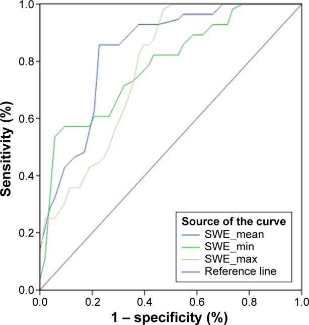
Receiver-operating characteristic curve analysis was performed to assess the diagnostic performance of 2D-SWE. Conventional US findings and 2D-SWE values were analyzed and compared between benign and malignant thyroid nodules. The mean values of SWE_mean, SWE_min, and SWE_max were 46.6±16.7, 26.2±9.5, and 73.6±18.1 kPa, respectively, in PTMC, which were significantly higher than those in benign tumors (27.8±12.4, 15.8±8.6, and 50.3±22.6 kPa, P<0.001). The optimal cut-off values of SWE_mean, SWE_min, and SWE_max for predicting malignancy were 34.5, 21.8, and 53.2 kPa, respectively. Taller than wide, micro-calcification, and SWE_mean were found to be independent risk factors for predicting PTMC. The overall sensitivity, specificity, accuracy, positive predictive value, and negative predictive value of combined conventional US features with 2D-SWE parameters were 95.7%, 94.5%, 94.9%, 89.8%, and 97.7%, respectively; these were superior to those of conventional US (89.1%, 90.1%, 89.9%, 82.0%, and 93.2%).

CONCLUSION

The study indicates that the quantitative parameters of 2D-SWE are an independent predictive factor for diagnosing PTMC, which could provide valuable information when conventional US cannot give determinate results.

摘要

目的

本研究旨在评估二维剪切波弹性成像(2D-SWE)对甲状腺微小乳头状癌(PTMC)的预测能力。

材料与方法

本研究纳入了118例患者的137个甲状腺结节(46个良性结节,91个恶性结节),这些患者在细针穿刺或手术前接受了传统超声(US)和2D-SWE检查。比较仅根据US检查结果以及US检查结果与2D-SWE联合使用时的诊断性能,并将其与病理结果相关联。

结果

进行了受试者操作特征曲线分析以评估2D-SWE的诊断性能。分析并比较了良性和恶性甲状腺结节的传统US检查结果和2D-SWE值。PTMC中SWE_mean、SWE_min和SWE_max的平均值分别为46.6±16.7、26.2±9.5和73.6±18.1kPa,显著高于良性肿瘤(27.8±12.4、15.8±8.6和50.3±22.6kPa,P<0.001)。预测恶性肿瘤的SWE_mean、SWE_min和SWE_max的最佳截断值分别为34.5、21.8和53.2kPa。纵横比大于1、微钙化和SWE_mean被发现是预测PTMC的独立危险因素。传统US特征与2D-SWE参数联合使用时的总体敏感性、特异性、准确性、阳性预测值和阴性预测值分别为95.7%、94.5%、94.9%、89.8%和97.7%;这些均优于传统US(89.1%、90.1%、89.9%、82.0%和93.2%)。

结论

该研究表明,2D-SWE的定量参数是诊断PTMC的独立预测因素,当传统US无法给出明确结果时,其可为诊断提供有价值的信息。

https://cdn.ncbi.nlm.nih.gov/pmc/blobs/dc15/4790519/3aa51e4b77c1/ott-9-1311Fig3.jpg
https://cdn.ncbi.nlm.nih.gov/pmc/blobs/dc15/4790519/f287177058b6/ott-9-1311Fig1.jpg
https://cdn.ncbi.nlm.nih.gov/pmc/blobs/dc15/4790519/d721509b3946/ott-9-1311Fig2.jpg
https://cdn.ncbi.nlm.nih.gov/pmc/blobs/dc15/4790519/3aa51e4b77c1/ott-9-1311Fig3.jpg
https://cdn.ncbi.nlm.nih.gov/pmc/blobs/dc15/4790519/f287177058b6/ott-9-1311Fig1.jpg
https://cdn.ncbi.nlm.nih.gov/pmc/blobs/dc15/4790519/d721509b3946/ott-9-1311Fig2.jpg
https://cdn.ncbi.nlm.nih.gov/pmc/blobs/dc15/4790519/3aa51e4b77c1/ott-9-1311Fig3.jpg

相似文献

1
Diagnostic value of two-dimensional shear wave elastography in papillary thyroid microcarcinoma.二维剪切波弹性成像在甲状腺微小乳头状癌中的诊断价值
Onco Targets Ther. 2016 Mar 9;9:1311-7. doi: 10.2147/OTT.S98583. eCollection 2016.
2
Three-Dimensional Shear Wave Elastography for Differentiating Benign From Malignant Thyroid Nodules.用于鉴别甲状腺良恶性结节的三维剪切波弹性成像
J Ultrasound Med. 2018 Jul;37(7):1777-1788. doi: 10.1002/jum.14531. Epub 2018 Jan 9.
3
Quantitative assessment of shear-wave ultrasound elastography in thyroid nodules: diagnostic performance for predicting malignancy.剪切波超声弹性成像在甲状腺结节中的定量评估:预测恶性肿瘤的诊断性能。
Eur Radiol. 2013 Sep;23(9):2532-7. doi: 10.1007/s00330-013-2847-5. Epub 2013 Apr 19.
4
Comparison of Strain Elastography, Shear Wave Elastography, and Conventional Ultrasound in Diagnosing Thyroid Nodules.应变弹性成像、剪切波弹性成像与传统超声在诊断甲状腺结节中的比较
J Med Ultrasound. 2019 Jan-Mar;27(1):26-32. doi: 10.4103/JMU.JMU_46_18. Epub 2018 Jun 6.
5
Diagnostic Performance of a Combination of Shear Wave Elastography and B-Mode Ultrasonography in Differentiating Benign From Malignant Thyroid Nodules.剪切波弹性成像与B型超声联合应用对甲状腺良恶性结节的诊断效能
Clin Exp Otorhinolaryngol. 2020 May;13(2):186-193. doi: 10.21053/ceo.2019.01235. Epub 2020 Mar 11.
6
[Value of thyroid imaging reporting and data system and shear wave elastography for diagnosis of thyroid microcarcinoma].[甲状腺影像报告和数据系统及剪切波弹性成像在甲状腺微小癌诊断中的价值]
Lin Chuang Er Bi Yan Hou Tou Jing Wai Ke Za Zhi. 2016 Jan;30(2):118-22.
7
Comparisons and Combined Application of Two-Dimensional and Three-Dimensional Real-time Shear Wave Elastography in Diagnosis of Thyroid Nodules.二维与三维实时剪切波弹性成像在甲状腺结节诊断中的对比及联合应用
J Cancer. 2019 May 12;10(9):1975-1984. doi: 10.7150/jca.30135. eCollection 2019.
8
Diagnostic performance of a new two-dimensional shear wave elastography expression using siemens ultrasound system combined with ACR TI-RADS for classification of benign and malignant thyroid nodules: A prospective multi-center study.使用西门子超声系统结合美国放射学会(ACR)甲状腺影像报告和数据系统(TI-RADS)的新型二维剪切波弹性成像表达对甲状腺良恶性结节进行分类的诊断性能:一项前瞻性多中心研究。
Heliyon. 2023 Sep 27;9(10):e20472. doi: 10.1016/j.heliyon.2023.e20472. eCollection 2023 Oct.
9
First experience of comparisons between two different shear wave speed imaging systems in differentiating malignant from benign thyroid nodules.两种不同剪切波速度成像系统在鉴别甲状腺良恶性结节方面比较的首次经验。
Clin Hemorheol Microcirc. 2017;65(4):349-361. doi: 10.3233/CH-16197.
10
Two-dimensional shear wave elastography as promising diagnostic tool for predicting malignant thyroid nodules: a prospective single-centre experience.二维剪切波弹性成像作为预测甲状腺恶性结节的有前景的诊断工具:一项前瞻性单中心研究经验
Eur Radiol. 2015 Mar;25(3):624-34. doi: 10.1007/s00330-014-3455-8. Epub 2014 Oct 9.

引用本文的文献

1
Multimodal Ultrasound Radiomic Technology for Diagnosing Benign and Malignant Thyroid Nodules of Ti-Rads 4-5: A Multicenter Study.多模态超声放射组学技术在诊断 Ti-RADS 4-5 级甲状腺良恶性结节中的应用:一项多中心研究。
Sensors (Basel). 2024 Sep 25;24(19):6203. doi: 10.3390/s24196203.
2
Impact of Region-of-Interest Size on the Diagnostic Performance of Shear Wave Elastography in Differentiating Thyroid Nodules.感兴趣区大小对剪切波弹性成像鉴别甲状腺结节诊断性能的影响
Cancers (Basel). 2023 Oct 30;15(21):5214. doi: 10.3390/cancers15215214.
3
Diagnostic performance of a new two-dimensional shear wave elastography expression using siemens ultrasound system combined with ACR TI-RADS for classification of benign and malignant thyroid nodules: A prospective multi-center study.

本文引用的文献

1
Quantitative comparison of transient elastography (TE), shear wave elastography (SWE) and liver biopsy results of patients with chronic liver disease.慢性肝病患者瞬时弹性成像(TE)、剪切波弹性成像(SWE)与肝活检结果的定量比较。
J Phys Ther Sci. 2015 Aug;27(8):2465-8. doi: 10.1589/jpts.27.2465. Epub 2015 Aug 21.
2
Correlation between quantitative shear wave elastography and pathologic structures of thyroid lesions.甲状腺病变的定量剪切波弹性成像与病理结构之间的相关性
Ultrasound Med Biol. 2015 Sep;41(9):2326-32. doi: 10.1016/j.ultrasmedbio.2015.05.001. Epub 2015 Jun 6.
3
Role of shear-wave elastography (SWE) in complex cystic and solid breast lesions in comparison with conventional ultrasound.
使用西门子超声系统结合美国放射学会(ACR)甲状腺影像报告和数据系统(TI-RADS)的新型二维剪切波弹性成像表达对甲状腺良恶性结节进行分类的诊断性能:一项前瞻性多中心研究。
Heliyon. 2023 Sep 27;9(10):e20472. doi: 10.1016/j.heliyon.2023.e20472. eCollection 2023 Oct.
4
US-Elastography With Different Techniques for Thyroid Nodule Characterization: Systematic Review and Meta-analysis.采用不同技术的超声弹性成像对甲状腺结节的特征分析:系统评价与荟萃分析
Front Oncol. 2022 Mar 16;12:845549. doi: 10.3389/fonc.2022.845549. eCollection 2022.
5
SuperSonic shear imaging for the differentiation between benign and malignant thyroid nodules: a meta-analysis.超声剪切波弹性成像在鉴别甲状腺良恶性结节中的应用:一项荟萃分析
J Endocrinol Invest. 2022 Jul;45(7):1327-1339. doi: 10.1007/s40618-022-01765-y. Epub 2022 Mar 1.
6
Influence of lesion size on shear wave elastography in the diagnosis of benign and malignant thyroid nodules.病变大小对剪切波弹性成像诊断甲状腺良恶性结节的影响。
Sci Rep. 2021 Nov 3;11(1):21616. doi: 10.1038/s41598-021-01114-8.
7
Accuracy of shear wave elastography in characterization of thyroid nodules in children and adolescents.剪切波弹性成像在儿童及青少年甲状腺结节特征描述中的准确性
Insights Imaging. 2021 Sep 9;12(1):128. doi: 10.1186/s13244-021-01074-7.
8
Evaluation of thyroid nodules by shear wave elastography: a review of current knowledge.剪切波弹性成像评估甲状腺结节:当前知识综述。
J Endocrinol Invest. 2021 Oct;44(10):2043-2056. doi: 10.1007/s40618-021-01570-z. Epub 2021 Apr 16.
9
The Role of Shear Wave Elastography in the Discrimination Between Malignant and Benign Thyroid Nodules.剪切波弹性成像在鉴别甲状腺良恶性结节中的作用
Acta Inform Med. 2020 Dec;28(4):248-253. doi: 10.5455/aim.2020.28.248-253.
10
Optimized algorithm in solid thyroid nodule elastography.实性甲状腺结节弹性成像中的优化算法
Oncol Lett. 2020 Nov;20(5):248. doi: 10.3892/ol.2020.12111. Epub 2020 Sep 16.
与传统超声相比,剪切波弹性成像(SWE)在乳腺复杂囊性和实性病变中的作用。
Eur J Radiol. 2015 Jul;84(7):1236-41. doi: 10.1016/j.ejrad.2015.04.005. Epub 2015 Apr 18.
4
Shear wave elastography of thyroid nodules for the prediction of malignancy in a large scale study.在一项大规模研究中,甲状腺结节的剪切波弹性成像用于预测恶性肿瘤。
Eur J Radiol. 2015 Mar;84(3):407-412. doi: 10.1016/j.ejrad.2014.11.019. Epub 2014 Nov 27.
5
Shear wave elastography versus real-time elastography on evaluation thyroid nodules: a preliminary study.剪切波弹性成像与实时弹性成像在评估甲状腺结节中的应用:一项初步研究。
Eur J Radiol. 2014 Jul;83(7):1135-1143. doi: 10.1016/j.ejrad.2014.02.024. Epub 2014 Mar 4.
6
Comparison of diagnostic value of conventional ultrasonography and shear wave elastography in the prediction of thyroid lesions malignancy.传统超声检查与剪切波弹性成像在预测甲状腺病变恶性程度方面的诊断价值比较。
PLoS One. 2013 Nov 29;8(11):e81532. doi: 10.1371/journal.pone.0081532. eCollection 2013.
7
Quantitative assessment of shear-wave ultrasound elastography in thyroid nodules: diagnostic performance for predicting malignancy.剪切波超声弹性成像在甲状腺结节中的定量评估:预测恶性肿瘤的诊断性能。
Eur Radiol. 2013 Sep;23(9):2532-7. doi: 10.1007/s00330-013-2847-5. Epub 2013 Apr 19.
8
Papillary thyroid microcarcinomas: a comparative study of the characteristics and risk factors at presentation in two cancer registries.甲状腺微小乳头状癌:两个癌症登记处中发病特征和危险因素的对比研究。
J Clin Endocrinol Metab. 2013 Apr;98(4):1427-34. doi: 10.1210/jc.2012-3728. Epub 2013 Mar 12.
9
Quantitative shear wave elastography as a prognostic implication of papillary thyroid carcinoma (PTC): elasticity index can predict extrathyroidal extension (ETE).定量剪切波弹性成像作为甲状腺乳头状癌 (PTC) 的预后意义:弹性指数可预测甲状腺外侵犯 (ETE)。
Ann Surg Oncol. 2013 Aug;20(8):2765-71. doi: 10.1245/s10434-013-2927-4. Epub 2013 Mar 6.
10
BRAF (V600E) mutation analysis on liquid-based cytology-processed aspiration biopsies predicts bilaterality and lymph node involvement in papillary thyroid microcarcinoma.液体细胞学处理的细针抽吸活检中 BRAF(V600E) 突变分析可预测甲状腺微小乳头状癌的双侧性和淋巴结受累。
Cancer Cytopathol. 2013 Jun;121(6):291-7. doi: 10.1002/cncy.21258. Epub 2012 Nov 28.