• 文献检索
  • 文档翻译
  • 深度研究
  • 学术资讯
  • Suppr Zotero 插件Zotero 插件
  • 邀请有礼
  • 套餐&价格
  • 历史记录
应用&插件
Suppr Zotero 插件Zotero 插件浏览器插件Mac 客户端Windows 客户端微信小程序
定价
高级版会员购买积分包购买API积分包
服务
文献检索文档翻译深度研究API 文档MCP 服务
关于我们
关于 Suppr公司介绍联系我们用户协议隐私条款
关注我们

Suppr 超能文献

核心技术专利:CN118964589B侵权必究
粤ICP备2023148730 号-1Suppr @ 2026

文献检索

告别复杂PubMed语法,用中文像聊天一样搜索,搜遍4000万医学文献。AI智能推荐,让科研检索更轻松。

立即免费搜索

文件翻译

保留排版,准确专业,支持PDF/Word/PPT等文件格式,支持 12+语言互译。

免费翻译文档

深度研究

AI帮你快速写综述,25分钟生成高质量综述,智能提取关键信息,辅助科研写作。

立即免费体验

利用磁共振成像和半自动处理技术评估健康受试者颈内静脉大小

Assessment of Internal Jugular Vein Size in Healthy Subjects with Magnetic Resonance and Semiautomatic Processing.

作者信息

Laganà M M, Pelizzari L, Scaccianoce E, Dipasquale O, Ricci C, Baglio F, Cecconi P, Baselli G

机构信息

IRCCS, Fondazione Don Carlo Gnocchi ONLUS, Via Capecelatro 66, 20148 Milan, Italy.

IRCCS, Fondazione Don Carlo Gnocchi ONLUS, Via Capecelatro 66, 20148 Milan, Italy; Department of Electronics, Information and Bioengineering, Politecnico di Milano, Piazza Leonardo da Vinci 32, 20133 Milan, Italy.

出版信息

Behav Neurol. 2016;2016:9717210. doi: 10.1155/2016/9717210. Epub 2016 Feb 29.

DOI:10.1155/2016/9717210
PMID:27034585
原文链接:https://pmc.ncbi.nlm.nih.gov/articles/PMC4789379/
Abstract

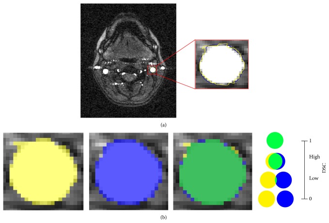
Background and Objectives. The hypothesized link between extracranial venous abnormalities and some neurological disorders awoke interest in the investigation of the internal jugular veins (IJVs). However, different IJV cross-sectional area (CSA) values are currently reported in literature. In this study, we introduced a semiautomatic method to measure and normalize the CSA and the degree of circularity (Circ) of IJVs along their whole length. Methods. Thirty-six healthy subjects (31.22 ± 9.29 years) were recruited and the 2D time-of-flight magnetic resonance venography was acquired with a 1.5 T Siemens scanner. The IJV were segmented on an axial slice, the contours were propagated in 3D. Then, IJV CSA and Circ were computed between the first and the seventh cervical levels (C1-C7) and normalized among subjects. Inter- and intrarater repeatability were assessed. Results. IJV CSA and Circ were significantly different among cervical levels (p < 0.001). A trend for side difference was observed for CSA (larger right IJV, p = 0.06), but not for Circ (p = 0.5). Excellent inter- and intrarater repeatability was obtained for all the measures. Conclusion. This study proposed a reliable semiautomatic method able to measure the IJV area and shape along C1-C7, and suitable for defining the normality thresholds for future clinical studies.

摘要

背景与目的。颅外静脉异常与某些神经系统疾病之间的假设联系引发了对颈内静脉(IJVs)研究的兴趣。然而,目前文献中报道的颈内静脉横截面积(CSA)值各不相同。在本研究中,我们引入了一种半自动方法来测量和标准化颈内静脉全长的CSA及圆度(Circ)。方法。招募了36名健康受试者(年龄31.22±9.29岁),使用1.5 T西门子扫描仪进行二维时间飞跃磁共振静脉成像。在轴位切片上分割颈内静脉,将轮廓在三维空间中传播。然后,计算第一至第七颈椎水平(C1 - C7)之间的颈内静脉CSA和Circ,并在受试者之间进行标准化。评估了评分者间和评分者内的重复性。结果。颈内静脉CSA和Circ在颈椎水平之间存在显著差异(p < 0.001)。观察到CSA存在侧别差异趋势(右侧颈内静脉较大,p = 0.06),但Circ不存在(p = 0.5)。所有测量均获得了出色的评分者间和评分者内重复性。结论。本研究提出了一种可靠的半自动方法,能够测量C1 - C7水平的颈内静脉面积和形状,适用于为未来临床研究确定正常阈值。

https://cdn.ncbi.nlm.nih.gov/pmc/blobs/e2d3/4789379/d622248eb62c/BN2016-9717210.003.jpg
https://cdn.ncbi.nlm.nih.gov/pmc/blobs/e2d3/4789379/2ad844614f98/BN2016-9717210.001.jpg
https://cdn.ncbi.nlm.nih.gov/pmc/blobs/e2d3/4789379/8e609492baca/BN2016-9717210.002.jpg
https://cdn.ncbi.nlm.nih.gov/pmc/blobs/e2d3/4789379/d622248eb62c/BN2016-9717210.003.jpg
https://cdn.ncbi.nlm.nih.gov/pmc/blobs/e2d3/4789379/2ad844614f98/BN2016-9717210.001.jpg
https://cdn.ncbi.nlm.nih.gov/pmc/blobs/e2d3/4789379/8e609492baca/BN2016-9717210.002.jpg
https://cdn.ncbi.nlm.nih.gov/pmc/blobs/e2d3/4789379/d622248eb62c/BN2016-9717210.003.jpg

相似文献

1
Assessment of Internal Jugular Vein Size in Healthy Subjects with Magnetic Resonance and Semiautomatic Processing.利用磁共振成像和半自动处理技术评估健康受试者颈内静脉大小
Behav Neurol. 2016;2016:9717210. doi: 10.1155/2016/9717210. Epub 2016 Feb 29.
2
Internal Jugular Vein Cross-Sectional Area Enlargement Is Associated with Aging in Healthy Individuals.健康个体的颈内静脉横截面积增大与衰老相关。
PLoS One. 2016 Feb 19;11(2):e0149532. doi: 10.1371/journal.pone.0149532. eCollection 2016.
3
Clinical significance of the cross-sectional area of the internal jugular vein.颈内静脉横截面积的临床意义。
J Cardiothorac Vasc Anesth. 2013 Aug;27(4):685-9. doi: 10.1053/j.jvca.2012.10.007. Epub 2013 Apr 30.
4
Magnetic Resonance Imaging of Internal Jugular Veins in Multiple Sclerosis: Interobserver Agreement and Comparison with Doppler Ultrasound Examination.多发性硬化症患者颈内静脉的磁共振成像:观察者间一致性及与多普勒超声检查的比较
Ann Vasc Surg. 2017 Jul;42:84-92. doi: 10.1016/j.avsg.2016.10.060. Epub 2017 Mar 11.
5
Internal Jugular Vein Cross-Sectional Area and Cerebrospinal Fluid Pulsatility in the Aqueduct of Sylvius: A Comparative Study between Healthy Subjects and Multiple Sclerosis Patients.颈内静脉横截面积与中脑导水管脑脊液搏动性:健康受试者与多发性硬化症患者的比较研究
PLoS One. 2016 May 2;11(5):e0153960. doi: 10.1371/journal.pone.0153960. eCollection 2016.
6
The effect of passive leg elevation and/or trendelenburg position on the cross-sectional area of the internal jugular vein in infants and young children undergoing surgery for congenital heart disease.被动抬腿和/或头高位对先天性心脏病手术患儿颈内静脉横截面积的影响。
Anesth Analg. 2013 Jan;116(1):178-84. doi: 10.1213/ANE.0b013e31826d2a89. Epub 2012 Dec 7.
7
An ultrasonographic technique to assess the jugular venous pulse: a proof of concept.一种评估颈静脉搏动的超声技术:概念验证
Ultrasound Med Biol. 2015 May;41(5):1334-41. doi: 10.1016/j.ultrasmedbio.2014.12.666. Epub 2015 Feb 18.
8
Internal jugular veins must be measured before catheterization.颈内静脉必须在插管前进行测量。
J Clin Anesth. 2015 Mar;27(2):129-31. doi: 10.1016/j.jclinane.2014.09.010. Epub 2014 Nov 24.
9
Neck Vessel Cross-Sectional Area Measured with MRI: Scan-Rescan Reproducibility for Longitudinal Evaluations.通过MRI测量的颈部血管横截面积:纵向评估的扫描-再扫描可重复性
J Neuroimaging. 2018 Jan;28(1):48-56. doi: 10.1111/jon.12488. Epub 2017 Dec 4.
10
Clinical Applicability of Assessment of Jugular Flow over the Individual Cardiac Cycle Compared with Current Ultrasound Methodology.与当前超声方法相比,评估个体心动周期中颈静脉血流的临床适用性。
Ultrasound Med Biol. 2016 Aug;42(8):1750-63. doi: 10.1016/j.ultrasmedbio.2016.03.002. Epub 2016 Apr 20.

引用本文的文献

1
Ultrasound Evaluation of Internal Jugular Venous Insufficiency and Its Association with Cognitive Decline.颈内静脉功能不全的超声评估及其与认知衰退的关联
Diagnostics (Basel). 2025 Jun 4;15(11):1427. doi: 10.3390/diagnostics15111427.
2
Connecting the dots: Linking superior ophthalmic vein and internal jugular vein diameter to carotid cavernous fistula type and location.理清头绪:将眼上静脉和颈内静脉直径与海绵窦瘘的类型及位置相联系。
Surg Neurol Int. 2024 Oct 18;15:377. doi: 10.25259/SNI_601_2024. eCollection 2024.
3
Five-Year Longitudinal Study of Neck Vessel Cross-Sectional Area in Multiple Sclerosis.

本文引用的文献

1
An anatomy-based lumped parameter model of cerebrospinal venous circulation: can an extracranial anatomical change impact intracranial hemodynamics?基于解剖学的脑脊液静脉循环集总参数模型:颅外解剖结构变化会影响颅内血流动力学吗?
BMC Neurol. 2015 Jun 23;15:95. doi: 10.1186/s12883-015-0352-y.
2
Recommendations for multimodal noninvasive and invasive screening for detection of extracranial venous abnormalities indicative of chronic cerebrospinal venous insufficiency: a position statement of the International Society for Neurovascular Disease.检测提示慢性脑脊髓静脉功能不全的颅外静脉异常的多模式无创和有创筛查建议:国际神经血管疾病协会立场声明
J Vasc Interv Radiol. 2014 Nov;25(11):1785-94.e17. doi: 10.1016/j.jvir.2014.07.024. Epub 2014 Sep 23.
3
多发性硬化症患者颈部血管横截面积的 5 年纵向研究。
AJNR Am J Neuroradiol. 2018 Sep;39(9):1703-1709. doi: 10.3174/ajnr.A5738. Epub 2018 Jul 26.
4
Neck Vessel Cross-Sectional Area Measured with MRI: Scan-Rescan Reproducibility for Longitudinal Evaluations.通过MRI测量的颈部血管横截面积:纵向评估的扫描-再扫描可重复性
J Neuroimaging. 2018 Jan;28(1):48-56. doi: 10.1111/jon.12488. Epub 2017 Dec 4.
5
Jugular Anomalies in Multiple Sclerosis Are Associated with Increased Collateral Venous Flow.多发性硬化症中的颈静脉异常与侧支静脉血流增加有关。
AJNR Am J Neuroradiol. 2017 Aug;38(8):1617-1622. doi: 10.3174/ajnr.A5219. Epub 2017 May 25.
MR venography in children and adolescents with multiple sclerosis does not show increased prevalence of extracranial veins anomalies.磁共振静脉造影显示儿童和青少年多发性硬化症患者颅外静脉异常的发生率并未增加。
Eur J Paediatr Neurol. 2014 Mar;18(2):218-22. doi: 10.1016/j.ejpn.2013.11.013. Epub 2013 Dec 25.
4
Internal jugular venous abnormalities in transient monocular blindness.暂时性单眼盲中的颈内静脉异常。
BMC Neurol. 2013 Jul 22;13:94. doi: 10.1186/1471-2377-13-94.
5
A comparative study of magnetic resonance venography techniques for the evaluation of the internal jugular veins in multiple sclerosis patients.磁共振静脉造影技术在多发性硬化症患者颈内静脉评估中的对比研究。
Magn Reson Imaging. 2013 Dec;31(10):1668-76. doi: 10.1016/j.mri.2013.05.012. Epub 2013 Jul 12.
6
Differences between internal jugular vein and vertebral vein flow examined in real time with the use of multigate ultrasound color Doppler.应用多门控彩色超声多普勒实时检测颈内静脉与椎静脉血流的差异。
AJNR Am J Neuroradiol. 2013 Oct;34(10):2000-4. doi: 10.3174/ajnr.A3557. Epub 2013 May 30.
7
Clinical significance of the cross-sectional area of the internal jugular vein.颈内静脉横截面积的临床意义。
J Cardiothorac Vasc Anesth. 2013 Aug;27(4):685-9. doi: 10.1053/j.jvca.2012.10.007. Epub 2013 Apr 30.
8
Screening for chronic cerebrospinal venous insufficiency (CCSVI) using ultrasound: recommendations for a protocol.使用超声筛查慢性脑脊髓静脉功能不全(CCSVI):方案建议
Funct Neurol. 2011 Oct-Dec;26(4):229-48.
9
Multiple sclerosis: cerebral circulation time.多发性硬化症:脑循环时间。
Radiology. 2012 Mar;262(3):947-55. doi: 10.1148/radiol.11111239.
10
Chronic cerebrospinal venous insufficiency in multiple sclerosis: diagnostic, pathogenetic, clinical and treatment perspectives.多发性硬化症中的慢性脑脊髓静脉功能不全:诊断、发病机制、临床和治疗观点。
Expert Rev Neurother. 2011 Sep;11(9):1277-94. doi: 10.1586/ern.11.117.