Martin Anna, Kronbichler Martin, Richlan Fabio
Centre for Cognitive Neuroscience, University of Salzburg, Hellbrunnerstr. 34, Salzburg, 5020, Austria.
Department of Psychology, University of Salzburg, Hellbrunnerstr. 34, Salzburg, 5020, Austria.
Hum Brain Mapp. 2016 Jul;37(7):2676-99. doi: 10.1002/hbm.23202. Epub 2016 Apr 7.
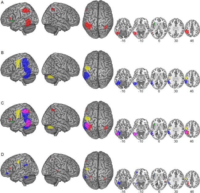
We used coordinate-based meta-analysis to objectively quantify commonalities and differences of dyslexic functional brain abnormalities between alphabetic languages differing in orthographic depth. Specifically, we compared foci of under- and overactivation in dyslexic readers relative to nonimpaired readers reported in 14 studies in deep orthographies (DO: English) and in 14 studies in shallow orthographies (SO: Dutch, German, Italian, Swedish). The separate meta-analyses of the two sets of studies showed universal reading-related dyslexic underactivation in the left occipitotemporal cortex (including the visual word form area (VWFA)). The direct statistical comparison revealed higher convergence of underactivation for DO compared with SO in bilateral inferior parietal regions, but this abnormality disappeared when foci resulting from stronger dyslexic task-negative activation (i.e., deactivation relative to baseline) were excluded. Higher convergence of underactivation for DO compared with SO was further identified in the left inferior frontal gyrus (IFG) pars triangularis, left precuneus, and right superior temporal gyrus, together with higher convergence of overactivation in the left anterior insula. Higher convergence of underactivation for SO compared with DO was found in the left fusiform gyrus, left temporoparietal cortex, left IFG pars orbitalis, and left frontal operculum, together with higher convergence of overactivation in the left precentral gyrus. Taken together, the findings support the notion of a biological unity of dyslexia, with additional orthography-specific abnormalities and presumably different compensatory mechanisms. The results are discussed in relation to current functional neuroanatomical models of developmental dyslexia. Hum Brain Mapp 37:2676-2699, 2016. © 2016 The Authors Human Brain Mapping Published by Wiley Periodicals, Inc.
我们采用基于坐标的元分析,客观地量化了正字法深度不同的字母语言之间诵读困难功能性脑区异常的共性与差异。具体而言,我们比较了14项关于深度正字法(DO:英语)研究和14项关于浅度正字法(SO:荷兰语、德语、意大利语、瑞典语)研究中,诵读困难读者相对于非受损读者的激活不足和激活过度的脑区。两组研究的单独元分析均显示,在左侧枕颞叶皮层(包括视觉词形区(VWFA))存在与阅读相关的普遍诵读困难激活不足。直接的统计比较显示,与浅度正字法相比,深度正字法在双侧顶下叶区域激活不足的汇聚程度更高,但当排除因诵读困难任务负激活更强(即相对于基线的失活)导致的脑区后,这种异常消失。与浅度正字法相比,深度正字法在左侧额下回(IFG)三角部、左侧楔前叶和右侧颞上回的激活不足汇聚程度更高,同时在左侧前岛叶的激活过度汇聚程度也更高。与深度正字法相比,浅度正字法在左侧梭状回、左侧颞顶叶皮层、左侧IFG眶部和左侧额盖的激活不足汇聚程度更高,同时在左侧中央前回的激活过度汇聚程度也更高。综上所述,这些发现支持了诵读困难具有生物学统一性的观点,同时存在额外的正字法特异性异常以及可能不同的补偿机制。我们结合当前发育性诵读困难的功能性神经解剖模型对结果进行了讨论。《人类大脑图谱》37:2676 - 2699, 2016。© 2016作者《人类大脑图谱》由威利期刊公司出版。