Wang Jinlin, Wu Xiaoli, Yin Ping, Guo Qiaozhen, Hou Wei, Li Yawen, Wang Yun, Cheng Bin
Department of Gastroenterology and Hepatology, Tongji Hospital, Tongji Medical College, Huazhong University of Science and Technology, Wuhan, 430030, China.
School of Public Health, Tongji Medical College, Huazhong University of Science and Technology, Wuhan, 430030, China.
Trials. 2016 Apr 12;17:198. doi: 10.1186/s13063-016-1316-2.
Linear endoscopic ultrasonography (EUS) allows the visualization, identification, and characterization of the extent of lesions of the gastrointestinal (GI) tract and adjacent structures. EUS-guided fine-needle aspiration (EUS-FNA) facilitates a more accurate diagnosis of mediastinal, intra-abdominal, and pancreatic lesions through the collection of the cytological material under direct visualization. Recent reports suggest that histological samples can be obtained by EUS-FNA with a reverse, bevel-tipped needle (the ProCore needle) to collect the core samples (fine needle biopsy, FNB), thereby adding a new dimension to the diagnostic usefulness of this technique. Certain neoplasms, such as lymphoma and stromal tumors, can be assessed by EUS-FNB to confirm the diagnosis. Here, we aimed to carry out a prospective, multicenter, single-blind, randomized, controlled trial to compare EUS-FNB and EUS-FNA.
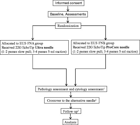
METHODS/DESIGN: A total of 408 patients will be enrolled from five endoscopic centers. Patients will be divided into two groups: (1) group A, which is the EUS regular needle group (EUS-FNA) and (2) group B, which is the EUS ProCore needle group (EUS-FNB). Patients in group A will be examined with a 22G EchoTip Ultra needle, and patients in group B, with a 22G EchoTip ProCore needle. For all included patients, four EUS-guided passes will be made in each lesion. In the first and second pass, a slow-pull suction method of the stylet will be done. The third and fourth pass will use manual suction of 5 cc. The primary objective is to compare the diagnostic yield of malignancy by EUS-FNA versus EUS-FNB.
The trial will compare samples obtained by EUS-FNA versus EUS-FNB for the diagnostic yield of solid lesions. The efficacy of these two sampling methods will be assessed on various lesions, which may provide insights into developing practice guidelines for their future indications.
Clinical Trials.gov, NCT02327065 .
线性超声内镜检查(EUS)能够对胃肠道(GI)及其相邻结构的病变范围进行可视化、识别和特征描述。EUS引导下细针穿刺抽吸术(EUS-FNA)可在直视下收集细胞学材料,有助于更准确地诊断纵隔、腹腔内及胰腺病变。近期报告表明,使用反向斜面尖端针(ProCore针)通过EUS-FNA可获取组织学样本以收集核心样本(细针活检,FNB),从而为该技术的诊断效用增添了新的维度。某些肿瘤,如淋巴瘤和间质瘤,可通过EUS-FNB进行评估以确诊。在此,我们旨在开展一项前瞻性、多中心、单盲、随机对照试验,以比较EUS-FNB和EUS-FNA。
方法/设计:将从五个内镜中心招募408例患者。患者将被分为两组:(1)A组,即EUS常规针组(EUS-FNA);(2)B组,即EUS ProCore针组(EUS-FNB)。A组患者将使用22G EchoTip Ultra针进行检查,B组患者将使用22G EchoTip ProCore针进行检查。对于所有纳入的患者,每个病变部位将进行4次EUS引导穿刺。在第一次和第二次穿刺中,将采用缓慢拔出针芯的抽吸方法。第三次和第四次穿刺将采用5毫升手动抽吸。主要目的是比较EUS-FNA与EUS-FNB对恶性肿瘤的诊断率。
该试验将比较EUS-FNA与EUS-FNB获取的样本对实体病变的诊断率。将在各种病变上评估这两种采样方法的疗效,这可能为制定其未来适应证的实践指南提供见解。
ClinicalTrials.gov,NCT02327065 。