Becerra Sandra C, Roy Daniel C, Sanchez Carlos J, Christy Robert J, Burmeister David M
Extremity Trauma and Regenerative Medicine Task Area, United States Army Institute of Surgical Research, 3650 Chambers Pass, JBSA Fort Sam Houston, TX, 78234, USA.
BMC Res Notes. 2016 Apr 12;9:216. doi: 10.1186/s13104-016-1902-0.
Bacterial infections are a common clinical problem in both acute and chronic wounds. With growing concerns over antibiotic resistance, treatment of bacterial infections should only occur after positive diagnosis. Currently, diagnosis is delayed due to lengthy culturing methods which may also fail to identify the presence of bacteria. While newer costly bacterial identification methods are being explored, a simple and inexpensive diagnostic tool would aid in immediate and accurate treatments for bacterial infections. Histologically, hematoxylin and eosin (H&E) and Gram stains have been employed, but are far from optimal when analyzing tissue samples due to non-specific staining. The goal of the current study was to develop a modification of the Gram stain that enhances the contrast between bacteria and host tissue.
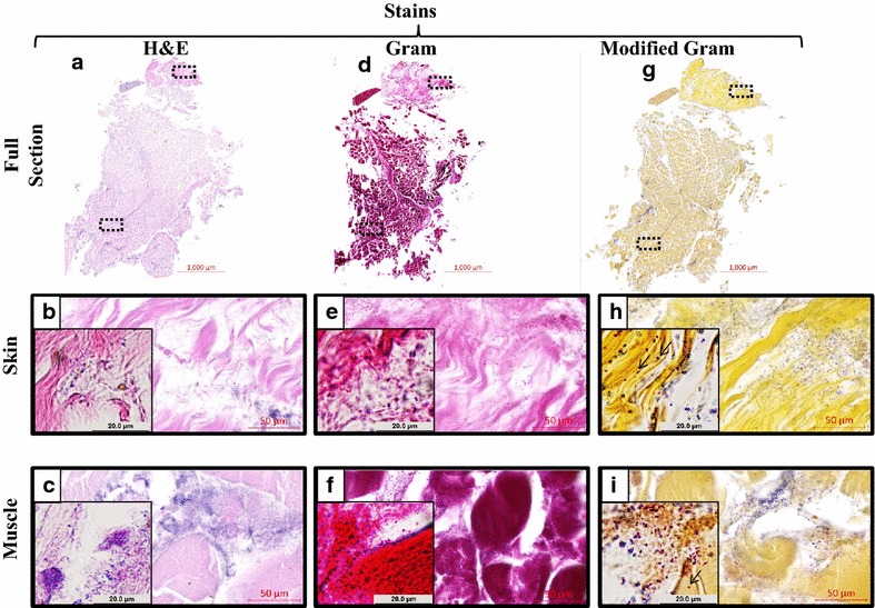
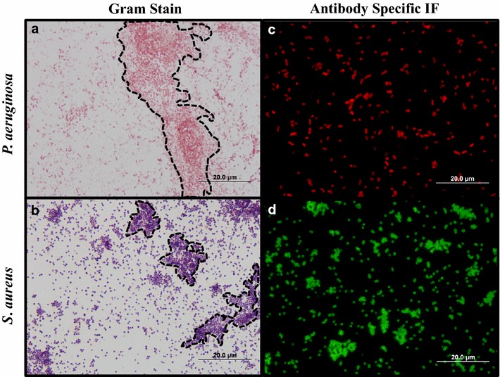
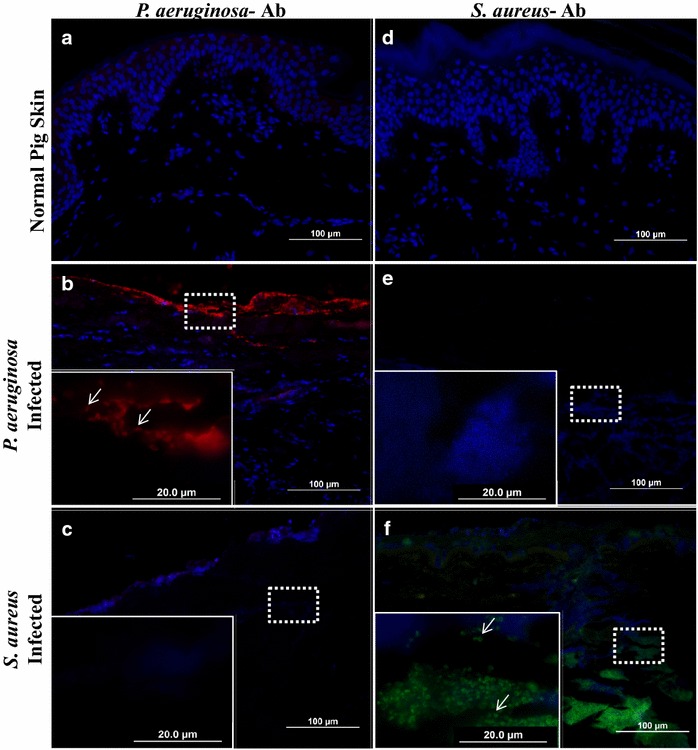
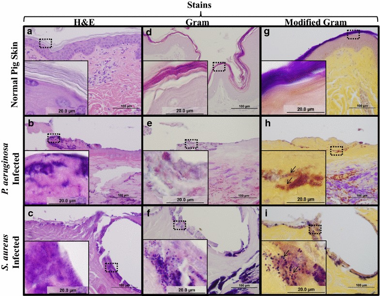
A modified Gram stain was developed and tested as an alternative to Gram stain that improves the contrast between Gram positive bacteria, Gram negative bacteria and host tissue. Initially, clinically relevant strains of Pseudomonas aeruginosa and Staphylococcus aureus were visualized in vitro and in biopsies of infected, porcine burns using routine Gram stain, and immunohistochemistry techniques involving bacterial strain-specific fluorescent antibodies as validation tools. H&E and Gram stain of serial biopsy sections were then compared to a modification of the Gram stain incorporating a counterstain that highlights collagen found in tissue. The modified Gram stain clearly identified both Gram positive and Gram negative bacteria, and when compared to H&E or Gram stain alone provided excellent contrast between bacteria and non-viable burn eschar. Moreover, when applied to surgical biopsies from patients that underwent burn debridement this technique was able to clearly detect bacterial morphology within host tissue.
We describe a modification of the Gram stain that provides improved contrast of Gram positive and Gram negative microorganisms within host tissue. The samples used in this study demonstrate that this staining technique has laboratory and clinical applicability. This modification only adds minutes to traditional Gram stain with reusable reagents, and results in a cost- and time-efficient technique for identifying bacteria in any clinical biopsy containing connective tissue.
细菌感染是急性和慢性伤口常见的临床问题。随着对抗生素耐药性的日益关注,细菌感染的治疗应仅在确诊后进行。目前,由于培养方法耗时较长,诊断往往延迟,而且可能无法识别细菌的存在。虽然正在探索更新的、成本更高的细菌鉴定方法,但一种简单且廉价的诊断工具将有助于对细菌感染进行即时和准确的治疗。在组织学上,苏木精和伊红(H&E)染色以及革兰氏染色已被采用,但在分析组织样本时,由于非特异性染色,它们远非最佳选择。本研究的目的是开发一种改良的革兰氏染色方法,以增强细菌与宿主组织之间的对比度。
开发并测试了一种改良的革兰氏染色方法,作为革兰氏染色的替代方法,该方法可改善革兰氏阳性菌、革兰氏阴性菌与宿主组织之间的对比度。最初,使用常规革兰氏染色以及涉及细菌菌株特异性荧光抗体的免疫组织化学技术作为验证工具,在体外以及感染的猪烧伤活检组织中观察了临床相关的铜绿假单胞菌和金黄色葡萄球菌菌株。然后,将连续活检切片的H&E染色和革兰氏染色与一种改良的革兰氏染色方法进行比较,该改良方法采用了一种复染剂,可突出组织中的胶原蛋白。改良的革兰氏染色能够清晰地识别革兰氏阳性菌和革兰氏阴性菌,与单独的H&E染色或革兰氏染色相比,在细菌与无活力的烧伤焦痂之间提供了出色的对比度。此外,当应用于接受烧伤清创术患者的手术活检组织时,该技术能够清晰地检测宿主组织内的细菌形态。
我们描述了一种改良的革兰氏染色方法,该方法可改善宿主组织内革兰氏阳性和革兰氏阴性微生物的对比度。本研究中使用的样本表明,这种染色技术具有实验室和临床适用性。这种改良仅在传统革兰氏染色的基础上增加了几分钟时间,且试剂可重复使用,从而形成了一种经济高效的技术,用于在任何含有结缔组织的临床活检中鉴定细菌。