• 文献检索
  • 文档翻译
  • 深度研究
  • 学术资讯
  • Suppr Zotero 插件Zotero 插件
  • 邀请有礼
  • 套餐&价格
  • 历史记录
应用&插件
Suppr Zotero 插件Zotero 插件浏览器插件Mac 客户端Windows 客户端微信小程序
定价
高级版会员购买积分包购买API积分包
服务
文献检索文档翻译深度研究API 文档MCP 服务
关于我们
关于 Suppr公司介绍联系我们用户协议隐私条款
关注我们

Suppr 超能文献

核心技术专利:CN118964589B侵权必究
粤ICP备2023148730 号-1Suppr @ 2026

文献检索

告别复杂PubMed语法,用中文像聊天一样搜索,搜遍4000万医学文献。AI智能推荐,让科研检索更轻松。

立即免费搜索

文件翻译

保留排版,准确专业,支持PDF/Word/PPT等文件格式,支持 12+语言互译。

免费翻译文档

深度研究

AI帮你快速写综述,25分钟生成高质量综述,智能提取关键信息,辅助科研写作。

立即免费体验

用于X射线计算机断层扫描的韧带和肌腱的最佳造影剂染色

Optimal Contrast Agent Staining of Ligaments and Tendons for X-Ray Computed Tomography.

作者信息

Balint Richard, Lowe Tristan, Shearer Tom

机构信息

School of Materials, University of Manchester, Manchester, United Kingdom.

Henry Moseley X-ray Imaging Facility, School of Materials, University of Manchester, Manchester, United Kingdom.

出版信息

PLoS One. 2016 Apr 14;11(4):e0153552. doi: 10.1371/journal.pone.0153552. eCollection 2016.

DOI:10.1371/journal.pone.0153552
PMID:27078030
原文链接:https://pmc.ncbi.nlm.nih.gov/articles/PMC4831740/
Abstract

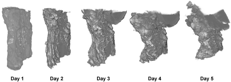
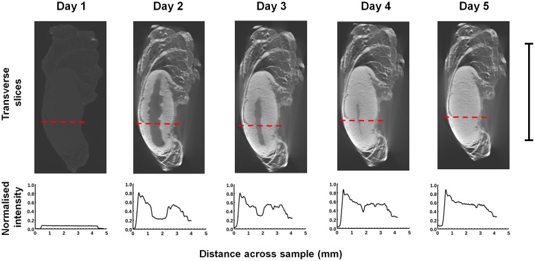
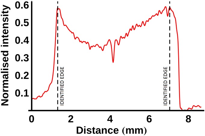
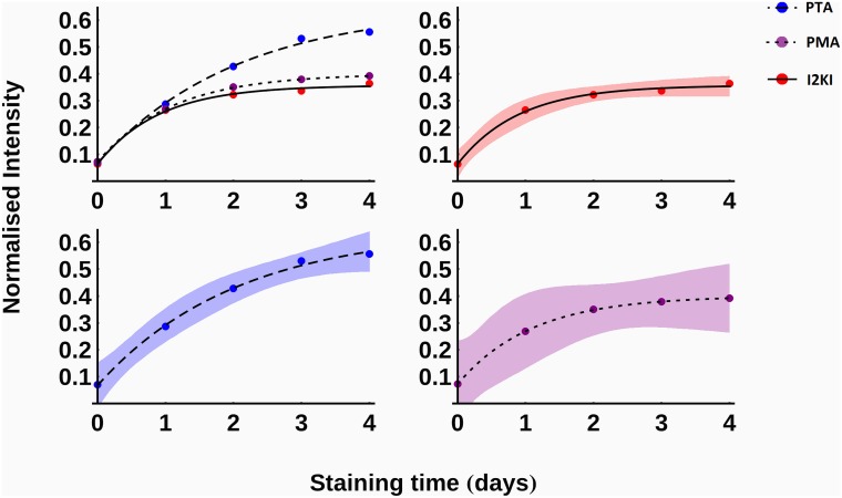
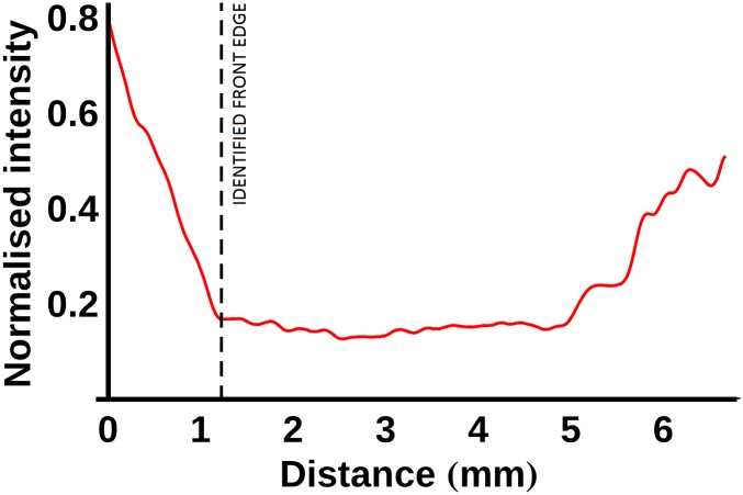
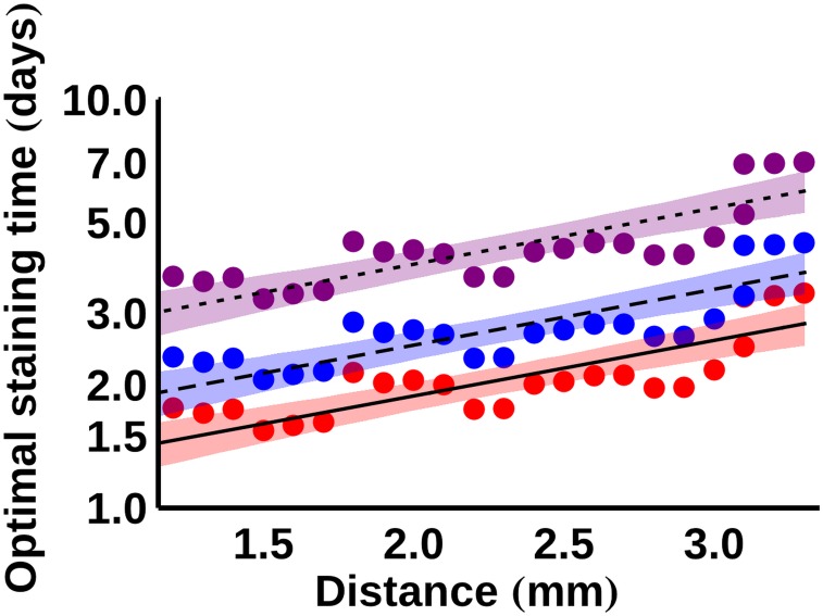
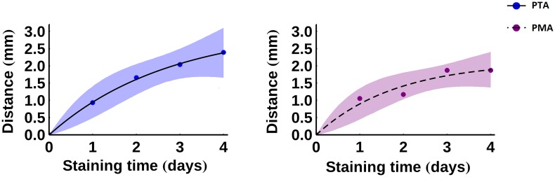
X-ray computed tomography has become an important tool for studying the microstructures of biological soft tissues, such as ligaments and tendons. Due to the low X-ray attenuation of such tissues, chemical contrast agents are often necessary to enhance contrast during scanning. In this article, the effects of using three different contrast agents--iodine potassium iodide solution, phosphotungstic acid and phosphomolybdic acid--are evaluated and compared. Porcine anterior cruciate ligaments, patellar tendons, medial collateral ligaments and lateral collateral ligaments were used as the basis of the study. Three samples of each of the four ligament/tendon types were each assigned a different contrast agent (giving a total of twelve samples), and the progression of that agent through the tissue was monitored by performing a scan every day for a total period of five days (giving a total of sixty scans). Since the samples were unstained on day one, they had been stained for a total of four days by the time of the final scans. The relative contrast enhancement and tissue deformation were measured. It was observed that the iodine potassium iodide solution penetrated the samples fastest and caused the least sample shrinkage on average (although significant deformation was observed by the time of the final scans), whereas the phosphomolybdic acid caused the greatest sample shrinkage. Equations describing the observed behaviour of the contrast agents, which can be used to predict optimal staining times for ligament and tendon X-ray computed tomography, are presented.

摘要

X射线计算机断层扫描已成为研究生物软组织(如韧带和肌腱)微观结构的重要工具。由于此类组织的X射线衰减较低,在扫描过程中通常需要使用化学造影剂来增强对比度。在本文中,对使用三种不同造影剂——碘化钾碘溶液、磷钨酸和磷钼酸——的效果进行了评估和比较。研究以猪的前交叉韧带、髌腱、内侧副韧带和外侧副韧带为基础。四种韧带/肌腱类型中的每种各取三个样本,分别使用不同的造影剂(共十二个样本),并通过在总共五天的时间里每天进行一次扫描(共六十次扫描)来监测造影剂在组织中的渗透过程。由于样本在第一天未染色,到最后一次扫描时它们总共被染色了四天。测量了相对对比度增强和组织变形情况。观察到碘化钾碘溶液渗透样本的速度最快,平均导致的样本收缩最小(尽管在最后一次扫描时观察到了明显的变形),而磷钼酸导致的样本收缩最大。本文给出了描述造影剂观察行为的方程,这些方程可用于预测韧带和肌腱X射线计算机断层扫描的最佳染色时间。

https://cdn.ncbi.nlm.nih.gov/pmc/blobs/18a4/4831740/bcf2449f8ed7/pone.0153552.g009.jpg
https://cdn.ncbi.nlm.nih.gov/pmc/blobs/18a4/4831740/f3fcb09cf545/pone.0153552.g001.jpg
https://cdn.ncbi.nlm.nih.gov/pmc/blobs/18a4/4831740/d59695820a2d/pone.0153552.g002.jpg
https://cdn.ncbi.nlm.nih.gov/pmc/blobs/18a4/4831740/19bbd500c7a8/pone.0153552.g003.jpg
https://cdn.ncbi.nlm.nih.gov/pmc/blobs/18a4/4831740/4216fdfef651/pone.0153552.g004.jpg
https://cdn.ncbi.nlm.nih.gov/pmc/blobs/18a4/4831740/ef033db0e966/pone.0153552.g005.jpg
https://cdn.ncbi.nlm.nih.gov/pmc/blobs/18a4/4831740/98ad0c1c1b96/pone.0153552.g006.jpg
https://cdn.ncbi.nlm.nih.gov/pmc/blobs/18a4/4831740/cdcedc45d65c/pone.0153552.g007.jpg
https://cdn.ncbi.nlm.nih.gov/pmc/blobs/18a4/4831740/0b79c98e6ab2/pone.0153552.g008.jpg
https://cdn.ncbi.nlm.nih.gov/pmc/blobs/18a4/4831740/bcf2449f8ed7/pone.0153552.g009.jpg

相似文献

1
Optimal Contrast Agent Staining of Ligaments and Tendons for X-Ray Computed Tomography.用于X射线计算机断层扫描的韧带和肌腱的最佳造影剂染色
PLoS One. 2016 Apr 14;11(4):e0153552. doi: 10.1371/journal.pone.0153552. eCollection 2016.
2
Iodine potassium iodide improves the contrast-to-noise ratio of micro-computed tomography images of the human middle ear.碘化钾可提高人中耳微计算机断层扫描图像的对比度噪声比。
J Microsc. 2016 Dec;264(3):334-338. doi: 10.1111/jmi.12447. Epub 2016 Jul 21.
3
X-ray computed tomography of the anterior cruciate ligament and patellar tendon.前交叉韧带和髌腱的X线计算机断层扫描
Muscles Ligaments Tendons J. 2014 Jul 14;4(2):238-44. eCollection 2014 Apr.
4
Tissue-specific changes in size and shape of the ligaments and tendons of the porcine knee during post-natal growth.猪膝关节韧带和肌腱在出生后生长过程中的组织特异性大小和形状变化。
PLoS One. 2019 Oct 23;14(10):e0219637. doi: 10.1371/journal.pone.0219637. eCollection 2019.
5
Ultrasound of the elbow with emphasis on detailed assessment of ligaments, tendons, and nerves.肘部超声检查,重点是对韧带、肌腱和神经进行详细评估。
Eur J Radiol. 2015 Apr;84(4):671-81. doi: 10.1016/j.ejrad.2014.12.007. Epub 2014 Dec 19.
6
Tendons and ligaments are anatomically distinct but overlap in molecular and morphological features--a comparative study in an ovine model.肌腱和韧带在解剖学上有明显区别,但在分子和形态特征上存在重叠——一项在绵羊模型中的比较研究。
J Orthop Res. 2007 Apr;25(4):458-64. doi: 10.1002/jor.20218.
7
Differences in mRNA expression patterns between patellar tendons and anterior cruciate ligaments of immature pigs.未成熟猪髌腱与前交叉韧带之间mRNA表达模式的差异。
J Orthop Res. 1999 Nov;17(6):823-8. doi: 10.1002/jor.1100170606.
8
An exploratory study of contrast agents for soft tissue visualization by means of high resolution X-ray computed tomography imaging.高分辨率 X 射线计算机断层成像技术用于软组织可视化的对比剂的探索性研究。
J Microsc. 2013 Apr;250(1):21-31. doi: 10.1111/jmi.12013. Epub 2013 Feb 22.
9
Assessing the three-dimensional collagen network in soft tissues using contrast agents and high resolution micro-CT: Application to porcine iliac veins.使用造影剂和高分辨率微型计算机断层扫描评估软组织中的三维胶原网络:在猪髂静脉中的应用
C R Biol. 2015 Jul;338(7):425-33. doi: 10.1016/j.crvi.2015.04.009. Epub 2015 May 29.
10
How accurate and reproducible are the identification of cruciate and collateral ligament insertions using MRI?使用磁共振成像(MRI)对交叉韧带和侧副韧带附着点进行识别的准确性和可重复性如何?
Knee. 2016 Aug;23(4):575-81. doi: 10.1016/j.knee.2015.07.015. Epub 2016 May 28.

引用本文的文献

1
Evaluating the impact of contrast agents on micro and nano mechanics of soft-to-hard tissue interface.评估造影剂对软硬组织界面微观和纳米力学的影响。
Sci Rep. 2025 Aug 2;15(1):28267. doi: 10.1038/s41598-025-12729-6.
2
Understanding the Structure-Function Relationship through 3D Imaging and Biomechanical Analysis: A Novel Methodological Approach Applied to Anterior Cruciate Ligaments.通过三维成像和生物力学分析理解结构-功能关系:一种应用于前交叉韧带的新型方法学途径。
Biomimetics (Basel). 2024 Aug 8;9(8):477. doi: 10.3390/biomimetics9080477.
3
Full-Field Strain Measurements of the Muscle-Tendon Junction Using X-ray Computed Tomography and Digital Volume Correlation.

本文引用的文献

1
A new strain energy function for modelling ligaments and tendons whose fascicles have a helical arrangement of fibrils.一种用于模拟韧带和肌腱的新应变能函数,其束状结构具有螺旋排列的原纤维。
J Biomech. 2015 Sep 18;48(12):3017-25. doi: 10.1016/j.jbiomech.2015.07.032. Epub 2015 Aug 8.
2
A new strain energy function for the hyperelastic modelling of ligaments and tendons based on fascicle microstructure.一种基于束状微观结构的用于韧带和肌腱超弹性建模的新应变能函数。
J Biomech. 2015 Jan 21;48(2):290-7. doi: 10.1016/j.jbiomech.2014.11.031. Epub 2014 Nov 25.
3
X-ray computed tomography of the anterior cruciate ligament and patellar tendon.
使用X射线计算机断层扫描和数字体积相关技术对肌腱结合处进行全场应变测量
Bioengineering (Basel). 2024 Feb 6;11(2):162. doi: 10.3390/bioengineering11020162.
4
Visualization of rat tendon in three dimensions using micro-Computed Tomography.使用微型计算机断层扫描对大鼠肌腱进行三维可视化。
MethodsX. 2024 Jan 10;12:102565. doi: 10.1016/j.mex.2024.102565. eCollection 2024 Jun.
5
Optimal Agents for Visualizing Collagen Tissue Microarchitecture Using Contrast-Enhanced MicroCT.使用对比增强显微CT可视化胶原组织微观结构的最佳试剂
Pharmaceuticals (Basel). 2023 Dec 12;16(12):1719. doi: 10.3390/ph16121719.
6
Revealing the three-dimensional murine brain microstructure by contrast-enhanced computed tomography.通过对比增强计算机断层扫描揭示三维小鼠脑微观结构。
Front Neurosci. 2023 Mar 23;17:1141615. doi: 10.3389/fnins.2023.1141615. eCollection 2023.
7
Cryogenic contrast-enhanced microCT enables nondestructive 3D quantitative histopathology of soft biological tissues.低温对比增强 microCT 可实现软生物组织的无损 3D 定量组织病理学。
Nat Commun. 2022 Oct 20;13(1):6207. doi: 10.1038/s41467-022-34048-4.
8
Bayesian inference on a microstructural, hyperelastic model of tendon deformation.贝叶斯推断在肌腱变形的微观结构、超弹性模型上的应用。
J R Soc Interface. 2022 May;19(190):20220031. doi: 10.1098/rsif.2022.0031. Epub 2022 May 18.
9
Significance of the broad non-bony attachments of the anterior cruciate ligament on the tibial side.前交叉韧带胫骨侧广泛非骨性附着的意义。
Sci Rep. 2022 Apr 27;12(1):6844. doi: 10.1038/s41598-022-10806-8.
10
A quality optimization approach to image Achilles tendon microstructure by phase-contrast enhanced synchrotron micro-tomography.基于相衬增强同步辐射微层析成像的图像跟腱微结构的质量优化方法。
Sci Rep. 2021 Aug 27;11(1):17313. doi: 10.1038/s41598-021-96589-w.
前交叉韧带和髌腱的X线计算机断层扫描
Muscles Ligaments Tendons J. 2014 Jul 14;4(2):238-44. eCollection 2014 Apr.
4
Optimal iodine staining of cardiac tissue for X-ray computed tomography.用于X射线计算机断层扫描的心脏组织最佳碘染色。
PLoS One. 2014 Aug 29;9(8):e105552. doi: 10.1371/journal.pone.0105552. eCollection 2014.
5
Structural stabilization of tissue for embryo phenotyping using micro-CT with iodine staining.使用碘染色的 micro-CT 对胚胎表型进行组织结构稳定。
PLoS One. 2013 Dec 30;8(12):e84321. doi: 10.1371/journal.pone.0084321. eCollection 2013.
6
Concentration-dependent specimen shrinkage in iodine-enhanced microCT.碘增强 microCT 中浓度依赖性标本收缩。
J Anat. 2013 Aug;223(2):185-93. doi: 10.1111/joa.12068. Epub 2013 May 30.
7
An exploratory study of contrast agents for soft tissue visualization by means of high resolution X-ray computed tomography imaging.高分辨率 X 射线计算机断层成像技术用于软组织可视化的对比剂的探索性研究。
J Microsc. 2013 Apr;250(1):21-31. doi: 10.1111/jmi.12013. Epub 2013 Feb 22.
8
Stress free configuration of the human eye.人眼的无应力配置。
Med Eng Phys. 2013 Feb;35(2):211-6. doi: 10.1016/j.medengphy.2012.09.006. Epub 2012 Oct 5.
9
A novel 3D mouse embryo atlas based on micro-CT.基于微 CT 的新型 3D 鼠胚胎图谱
Development. 2012 Sep;139(17):3248-56. doi: 10.1242/dev.082016.
10
Application of micro-computed tomography with iodine staining to cardiac imaging, segmentation, and computational model development.碘染色微计算机断层扫描在心脏成像、分割和计算模型开发中的应用。
IEEE Trans Med Imaging. 2013 Jan;32(1):8-17. doi: 10.1109/TMI.2012.2209183. Epub 2012 Jul 17.