• 文献检索
  • 文档翻译
  • 深度研究
  • 学术资讯
  • Suppr Zotero 插件Zotero 插件
  • 邀请有礼
  • 套餐&价格
  • 历史记录
应用&插件
Suppr Zotero 插件Zotero 插件浏览器插件Mac 客户端Windows 客户端微信小程序
定价
高级版会员购买积分包购买API积分包
服务
文献检索文档翻译深度研究API 文档MCP 服务
关于我们
关于 Suppr公司介绍联系我们用户协议隐私条款
关注我们

Suppr 超能文献

核心技术专利:CN118964589B侵权必究
粤ICP备2023148730 号-1Suppr @ 2026

文献检索

告别复杂PubMed语法,用中文像聊天一样搜索,搜遍4000万医学文献。AI智能推荐,让科研检索更轻松。

立即免费搜索

文件翻译

保留排版,准确专业,支持PDF/Word/PPT等文件格式,支持 12+语言互译。

免费翻译文档

深度研究

AI帮你快速写综述,25分钟生成高质量综述,智能提取关键信息,辅助科研写作。

立即免费体验

上皮单层的前沿推进速度取决于其初始限制几何形状。

Advancing Edge Speeds of Epithelial Monolayers Depend on Their Initial Confining Geometry.

作者信息

Kollimada Somanna A, Kulkarni Ankur H, Ravan Aniket, Gundiah Namrata

机构信息

Department of Mechanical Engineering, Indian Institute of Science, Bangalore, India.

Department of Molecular Reproduction, Development and Genetics, Indian Institute of Science, Bangalore, India.

出版信息

PLoS One. 2016 Apr 14;11(4):e0153471. doi: 10.1371/journal.pone.0153471. eCollection 2016.

DOI:10.1371/journal.pone.0153471
PMID:27078632
原文链接:https://pmc.ncbi.nlm.nih.gov/articles/PMC4831833/
Abstract

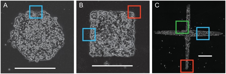
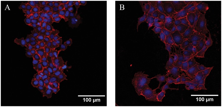
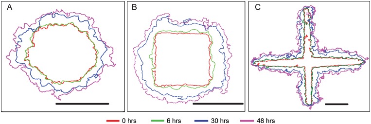
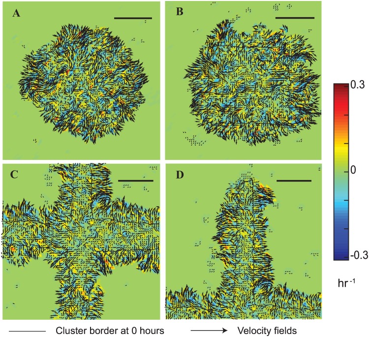
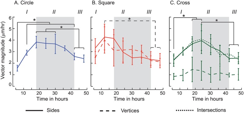
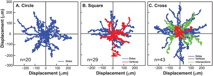
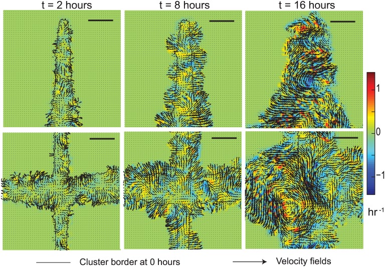
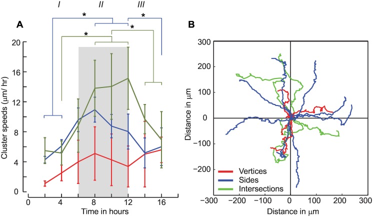
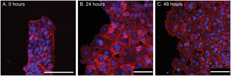
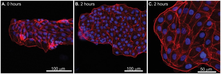
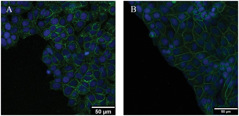
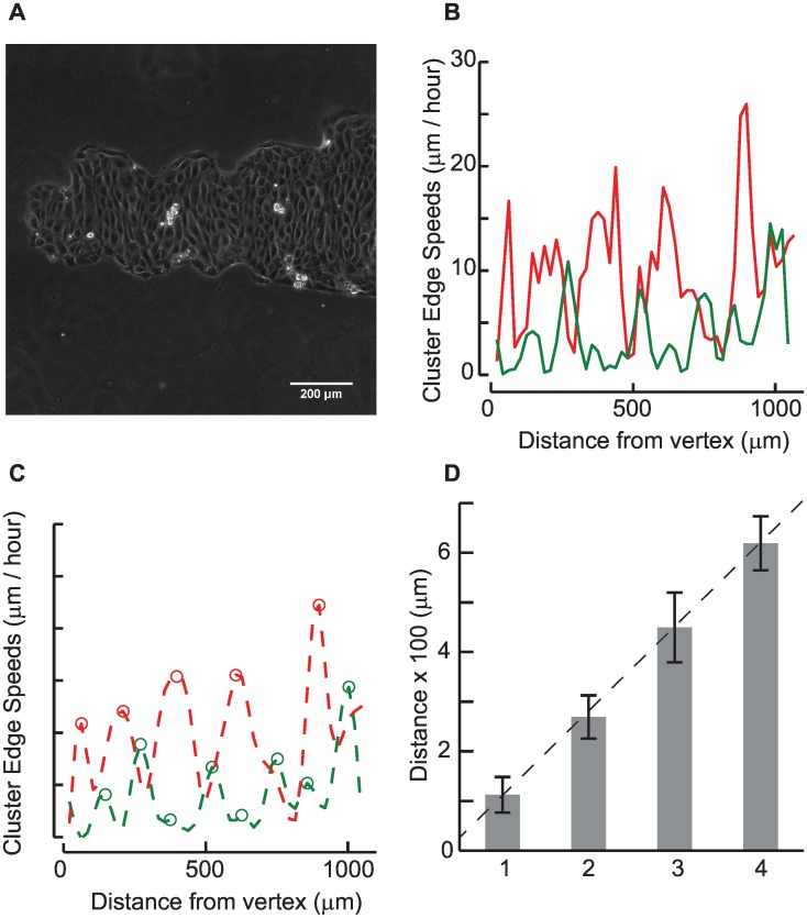
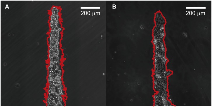
Collective cell migrations are essential in several physiological processes and are driven by both chemical and mechanical cues. The roles of substrate stiffness and confinement on collective migrations have been investigated in recent years, however few studies have addressed how geometric shapes influence collective cell migrations. Here, we address the hypothesis that the relative position of a cell within the confinement influences its motility. Monolayers of two types of epithelial cells--MCF7, a breast epithelial cancer cell line, and MDCK, a control epithelial cell line--were confined within circular, square, and cross-shaped stencils and their migration velocities were quantified upon release of the constraint using particle image velocimetry. The choice of stencil geometry allowed us to investigate individual cell motility within convex, straight and concave boundaries. Cells located in sharp, convex boundaries migrated at slower rates than those in concave or straight edges in both cell types. The overall cluster migration occurred in three phases: an initial linear increase with time, followed by a plateau region and a subsequent decrease in cluster speeds. An acto-myosin contractile ring, present in the MDCK but absent in MCF7 monolayer, was a prominent feature in the emergence of leader cells from the MDCK clusters which occurred every ~125 μm from the vertex of the cross. Further, coordinated cell movements displayed vorticity patterns in MDCK which were absent in MCF7 clusters. We also used cytoskeletal inhibitors to show the importance of acto-myosin bounding cables in collective migrations through translation of local movements to create long range coordinated movements and the creation of leader cells within ensembles. To our knowledge, this is the first demonstration of how bounding shapes influence long-term migratory behaviours of epithelial cell monolayers. These results are important for tissue engineering and may also enhance our understanding of cell movements during developmental patterning and cancer metastasis.

摘要

集体细胞迁移在多个生理过程中至关重要,且由化学和机械信号驱动。近年来,人们研究了底物硬度和限制条件对集体迁移的作用,然而,很少有研究探讨几何形状如何影响集体细胞迁移。在此,我们探讨这样一个假设:细胞在限制条件内的相对位置会影响其运动性。将两种上皮细胞单层——乳腺癌上皮细胞系MCF7和对照上皮细胞系MDCK——限制在圆形、方形和十字形模板内,并在解除限制后使用粒子图像测速法对其迁移速度进行量化。模板几何形状的选择使我们能够研究凸形、直线形和凹形边界内单个细胞的运动性。在两种细胞类型中,位于尖锐凸形边界的细胞迁移速度均低于凹形或直线形边界的细胞。整体集群迁移分为三个阶段:最初随时间呈线性增加,随后是平稳期,集群速度随后下降。在MDCK单层中存在而在MCF7单层中不存在的肌动蛋白-肌球蛋白收缩环,是MDCK集群中每隔约125μm从十字顶点出现的领头细胞的一个显著特征。此外,MDCK中的协调细胞运动显示出涡旋模式,而MCF7集群中则没有。我们还使用细胞骨架抑制剂来证明肌动蛋白-肌球蛋白结合索在集体迁移中的重要性,即通过局部运动的平移来产生长距离协调运动,并在群体中产生领头细胞。据我们所知,这是首次证明边界形状如何影响上皮细胞单层的长期迁移行为。这些结果对组织工程很重要,也可能增进我们对发育模式形成和癌症转移过程中细胞运动的理解。

https://cdn.ncbi.nlm.nih.gov/pmc/blobs/caed/4831833/0014db17fec3/pone.0153471.g014.jpg
https://cdn.ncbi.nlm.nih.gov/pmc/blobs/caed/4831833/3ff3e0d52a15/pone.0153471.g001.jpg
https://cdn.ncbi.nlm.nih.gov/pmc/blobs/caed/4831833/2c7882361850/pone.0153471.g002.jpg
https://cdn.ncbi.nlm.nih.gov/pmc/blobs/caed/4831833/331099af0874/pone.0153471.g003.jpg
https://cdn.ncbi.nlm.nih.gov/pmc/blobs/caed/4831833/0fa82c449b7a/pone.0153471.g004.jpg
https://cdn.ncbi.nlm.nih.gov/pmc/blobs/caed/4831833/8b349c2f66ef/pone.0153471.g005.jpg
https://cdn.ncbi.nlm.nih.gov/pmc/blobs/caed/4831833/e214152ffc2f/pone.0153471.g006.jpg
https://cdn.ncbi.nlm.nih.gov/pmc/blobs/caed/4831833/9ff9087ddae1/pone.0153471.g007.jpg
https://cdn.ncbi.nlm.nih.gov/pmc/blobs/caed/4831833/a8b08a9479c8/pone.0153471.g008.jpg
https://cdn.ncbi.nlm.nih.gov/pmc/blobs/caed/4831833/1f007eea709c/pone.0153471.g009.jpg
https://cdn.ncbi.nlm.nih.gov/pmc/blobs/caed/4831833/76a748216d4d/pone.0153471.g010.jpg
https://cdn.ncbi.nlm.nih.gov/pmc/blobs/caed/4831833/0c490b5a622d/pone.0153471.g011.jpg
https://cdn.ncbi.nlm.nih.gov/pmc/blobs/caed/4831833/efb708378eb5/pone.0153471.g012.jpg
https://cdn.ncbi.nlm.nih.gov/pmc/blobs/caed/4831833/ccc9cb58f9ce/pone.0153471.g013.jpg
https://cdn.ncbi.nlm.nih.gov/pmc/blobs/caed/4831833/0014db17fec3/pone.0153471.g014.jpg

相似文献

1
Advancing Edge Speeds of Epithelial Monolayers Depend on Their Initial Confining Geometry.上皮单层的前沿推进速度取决于其初始限制几何形状。
PLoS One. 2016 Apr 14;11(4):e0153471. doi: 10.1371/journal.pone.0153471. eCollection 2016.
2
Shear-induced damped oscillations in an epithelium depend on actomyosin contraction and E-cadherin cell adhesion.上皮细胞中的剪切诱导阻尼振荡依赖于肌动球蛋白收缩和 E-钙黏蛋白细胞黏附。
Elife. 2018 Nov 14;7:e39640. doi: 10.7554/eLife.39640.
3
Regulation of epithelial cell organization by tuning cell-substrate adhesion.通过调节细胞与底物的黏附来调控上皮细胞的组织架构。
Integr Biol (Camb). 2015 Oct;7(10):1228-41. doi: 10.1039/c5ib00196j. Epub 2015 Sep 24.
4
E-cadherin-dependent coordinated epithelial rotation on a two-dimensional discoidal pattern.E-钙黏蛋白依赖性的二维盘状模式下上皮细胞的协同旋转
Genes Cells. 2023 Mar;28(3):175-187. doi: 10.1111/gtc.13001. Epub 2022 Dec 30.
5
Substrate stiffness regulates cadherin-dependent collective migration through myosin-II contractility.基质硬度通过肌球蛋白 II 收缩调节钙黏着蛋白依赖性集体迁移。
J Cell Biol. 2012 Oct 29;199(3):545-63. doi: 10.1083/jcb.201207148. Epub 2012 Oct 22.
6
Guidance of collective cell migration by substrate geometry.基底几何形状对细胞集体迁移的导向作用。
Integr Biol (Camb). 2013 Aug;5(8):1026-35. doi: 10.1039/c3ib40054a.
7
Reduced adhesive ligand density in engineered extracellular matrices induces an epithelial-mesenchymal-like transition.工程化细胞外基质中黏附配体密度的降低会诱导上皮-间充质样转变。
Acta Biomater. 2016 Jul 15;39:106-113. doi: 10.1016/j.actbio.2016.05.006. Epub 2016 May 6.
8
Photoactivatable substrates for systematic study of the impact of an extracellular matrix ligand on appearance of leader cells in collective cell migration.用于系统研究细胞外基质配体对细胞集体迁移中先导细胞出现的影响的光激活底物。
Biomaterials. 2018 Jul;169:72-84. doi: 10.1016/j.biomaterials.2018.03.045. Epub 2018 Mar 27.
9
Delayed jamming-induced oscillatory migration patterns of epithelial collectives under long-range confinement.长程受限下延迟致阻塞诱导的上皮集体的振荡迁移模式。
Integr Biol (Camb). 2024 Jan 23;16. doi: 10.1093/intbio/zyae016.
10
Cellular Contraction and Polarization Drive Collective Cellular Motion.细胞收缩和极化驱动细胞集体运动。
Biophys J. 2016 Jun 21;110(12):2729-2738. doi: 10.1016/j.bpj.2016.05.019.

引用本文的文献

1
Extracellular matrix micropatterning technology for whole cell cryogenic electron microscopy studies.用于全细胞低温电子显微镜研究的细胞外基质微图案化技术
J Micromech Microeng. 2019 Nov;29(11). doi: 10.1088/1361-6439/ab419a. Epub 2019 Sep 26.
2
Switch-like enhancement of epithelial-mesenchymal transition by YAP through feedback regulation of WT1 and Rho-family GTPases.通过 YAP 对 WT1 和 Rho 家族 GTP 酶的反馈调节,实现上皮-间质转化的类开关增强。
Nat Commun. 2019 Jun 26;10(1):2797. doi: 10.1038/s41467-019-10729-5.
3
Controlling cell shape on hydrogels using lift-off protein patterning.

本文引用的文献

1
Modeling the finger instability in an expanding cell monolayer.模拟扩展细胞单层中的手指不稳定性。
Integr Biol (Camb). 2015 Oct;7(10):1218-27. doi: 10.1039/c5ib00092k. Epub 2015 Jun 22.
2
Leader cells regulate collective cell migration via Rac activation in the downstream signaling of integrin β1 and PI3K.引导细胞通过整合素β1和PI3K下游信号通路中的Rac激活来调节集体细胞迁移。
Sci Rep. 2015 Jan 7;5:7656. doi: 10.1038/srep07656.
3
Polarizing cytoskeletal tension to induce leader cell formation during collective cell migration.
利用剥离蛋白图案化技术在水凝胶上控制细胞形状。
PLoS One. 2018 Jan 3;13(1):e0189901. doi: 10.1371/journal.pone.0189901. eCollection 2018.
4
Leaders in collective migration: are front cells really endowed with a particular set of skills?集体迁移中的领导者:前端细胞真的具备一套特殊技能吗?
F1000Res. 2017 Oct 27;6:1899. doi: 10.12688/f1000research.11889.1. eCollection 2017.
5
Stereolithographic printing of ionically-crosslinked alginate hydrogels for degradable biomaterials and microfluidics.离子交联海藻酸钠水凝胶的立体光刻打印用于可降解生物材料和微流控。
Lab Chip. 2017 Oct 11;17(20):3474-3488. doi: 10.1039/c7lc00694b.
6
Deletion of the cytoplasmic domain of N-cadherin reduces, but does not eliminate, traction force-transmission.删除N-钙黏蛋白的细胞质结构域会降低但不会消除牵引力传递。
Biochem Biophys Res Commun. 2016 Sep 30;478(4):1640-6. doi: 10.1016/j.bbrc.2016.08.173. Epub 2016 Sep 3.
在集体细胞迁移过程中极化细胞骨架张力以诱导领头细胞形成。
Biointerphases. 2013 Dec;8(1):32. doi: 10.1186/1559-4106-8-32. Epub 2013 Nov 22.
4
Interplay of RhoA and mechanical forces in collective cell migration driven by leader cells.RhoA 与机械力在由先导细胞驱动的细胞集体迁移中的相互作用。
Nat Cell Biol. 2014 Mar;16(3):217-23. doi: 10.1038/ncb2917.
5
Guidance of collective cell migration by substrate geometry.基底几何形状对细胞集体迁移的导向作用。
Integr Biol (Camb). 2013 Aug;5(8):1026-35. doi: 10.1039/c3ib40054a.
6
Collective cell motion in an epithelial sheet can be quantitatively described by a stochastic interacting particle model.上皮片中的集体细胞运动可以通过随机相互作用粒子模型进行定量描述。
PLoS Comput Biol. 2013;9(3):e1002944. doi: 10.1371/journal.pcbi.1002944. Epub 2013 Mar 7.
7
E-cadherin-dependent stimulation of traction force at focal adhesions via the Src and PI3K signaling pathways.E-钙黏蛋白通过Src 和 PI3K 信号通路依赖性刺激黏着斑处的牵引力。
Biophys J. 2012 Jul 18;103(2):175-84. doi: 10.1016/j.bpj.2012.06.009. Epub 2012 Jul 17.
8
Collective cell migration: leadership, invasion and segregation.集体细胞迁移:领导、入侵和分离。
J R Soc Interface. 2012 Dec 7;9(77):3268-78. doi: 10.1098/rsif.2012.0448. Epub 2012 Jul 25.
9
Emerging modes of collective cell migration induced by geometrical constraints.几何约束诱导的细胞集体迁移的新兴模式。
Proc Natl Acad Sci U S A. 2012 Aug 7;109(32):12974-9. doi: 10.1073/pnas.1119313109. Epub 2012 Jul 19.
10
Collective and single cell behavior in epithelial contact inhibition.上皮细胞接触抑制中的群体和单细胞行为。
Proc Natl Acad Sci U S A. 2012 Jan 17;109(3):739-44. doi: 10.1073/pnas.1007809109. Epub 2012 Jan 6.