Khadka Deepak, Bhandari Sanjeeb, Bajimaya Sanyam, Thapa Raba, Paudyal Govinda, Pradhan Eli
Geta Eye Hospital, Dhangadi, Nepal.
Tilganga Institute of Ophthalmology, Kathmandu, Nepal.
BMC Ophthalmol. 2016 Apr 18;16:41. doi: 10.1186/s12886-016-0218-0.
Premacular subhyaloid hemorrhage results in a sudden profound loss of vision. Among the modalities for its treatment, Nd:YAG laser hyaloidotomy is a non invasive method enabling rapid drainage of the obstructed macular area and improved vision within days. This study was aimed to evaluate the efficacy, visual outcome and complications following Nd:YAG laser hyaloidotomy for premacular subhyaloid hemorrhage.
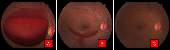
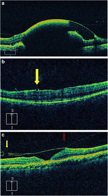
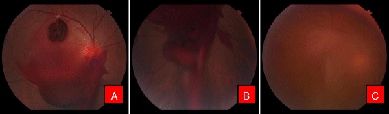
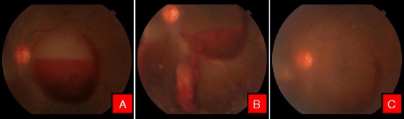
Patients with premacular subhyaloid hemorrhage of more than 3 disc diameters (DD) of various etiologies, attending Tilganga Institute of Ophthalmology, Nepal from August, 2014 to February, 2015, were included. A comprehensive ocular evaluation was conducted and fundus photographs were taken to measure the size of the subhyaloid hemorrhage. Optical coherence tomography (OCT) were performed before and after treatment and on subsequent follow up visits. Fundus fluorescence angiography was done whenever necessary. Q switched Nd:YAG laser was applied to create an opening in the posterior hyaloids membrane for draining subhyaloid hemorrhage. The main outcome measures were success rate in performing hyaloidotomy, drainage of subhyaloid blood into vitreous cavity and its resorption, improvement in visual acuity, need for further intervention and postoperative complications.
There were 21 eyes of 19 patients, 17(89.48%) male and 2(10.52%) female. In 3, premacular subhyaloid hemorrhage was bilateral. Mean age was 41.68 ± 17.08 years and a mean duration of symptoms 15.04 days. Mean pretreatment hemorrhage was 6.27DD. Nd:YAG laser hyaloidotomy was successful in 19 eyes(86.4%). In 2 patients, one each with Eales' disease and retinal vein occlusion the procedure was unsuccessful, necessitating pars plana vitrectomy, while in a case with proliferative diabetic retinopathy (PDR), vitrectomy was resorted for non clearing vitreous hemorrhage. Vision improved from a median of 3/60 pre-operatively to 6/6, at 6 months follow up. At 3 months, 2 patients with Eales' disease, one developed tractional detachment at macula while the other, an epiretinal membrane. No other complications were noted at 6 months.
Nd:YAG laser hyaloidotomy is an inexpensive, effective and a safe outpatient procedure for premacular subhyaloid hemorrhage, producing rapid drainage with restoration of visual function avoiding more invasive procedures and enabling early assessment of the underlying retina. The final visual prognosis however, rests on the underlying cause of the subhyaloid hemorrhage and any accompanying retinal changes.
黄斑前玻璃体下出血会导致视力突然严重丧失。在其治疗方法中,钕钇铝石榴石(Nd:YAG)激光玻璃体切割术是一种非侵入性方法,能够使阻塞的黄斑区域迅速引流,并在数天内改善视力。本研究旨在评估Nd:YAG激光玻璃体切割术治疗黄斑前玻璃体下出血的疗效、视觉结果及并发症。
纳入2014年8月至2015年2月在尼泊尔蒂尔冈加眼科研究所就诊的各种病因导致黄斑前玻璃体下出血超过3个视盘直径(DD)的患者。进行全面的眼部评估并拍摄眼底照片以测量玻璃体下出血的大小。在治疗前后及后续随访时进行光学相干断层扫描(OCT)。必要时进行眼底荧光血管造影。应用调Q Nd:YAG激光在玻璃体后膜上制造开口以引流玻璃体下出血。主要观察指标为玻璃体切割术的成功率、玻璃体下血液引流至玻璃体腔及其吸收情况、视力改善情况、进一步干预的必要性及术后并发症。
19例患者共21只眼,男性17例(89.48%),女性2例(10.52%)。3例黄斑前玻璃体下出血为双侧。平均年龄为41.68±17.08岁,平均症状持续时间为15.04天。术前平均出血面积为6.27DD。Nd:YAG激光玻璃体切割术在19只眼中成功(86.4%)。2例患者,1例患有伊尔斯病,1例患有视网膜静脉阻塞,手术未成功,需要行玻璃体切除术,而1例增殖性糖尿病视网膜病变(PDR)患者因玻璃体出血未清除而行玻璃体切除术。随访6个月时,视力从中位数术前的3/60提高到6/6。3个月时,2例伊尔斯病患者,1例发生黄斑部牵引性视网膜脱离,另1例发生视网膜前膜。6个月时未发现其他并发症。
Nd:YAG激光玻璃体切割术是一种治疗黄斑前玻璃体下出血的廉价、有效且安全的门诊手术,能迅速引流并恢复视觉功能,避免了更具侵入性的手术,且能早期评估潜在的视网膜情况。然而,最终的视觉预后取决于玻璃体下出血的潜在病因及任何伴随的视网膜变化。