Sonekatsu Mayumi, Taniguchi Wataru, Yamanaka Manabu, Nishio Naoko, Tsutsui Shunji, Yamada Hiroshi, Yoshida Munehito, Nakatsuka Terumasa
Department of Orthopaedic Surgery, Wakayama Medical University, Wakayama, Japan.
Department of Orthopaedic Surgery, Wakayama Medical University, Wakayama, Japan
Mol Pain. 2016 Apr 18;12. doi: 10.1177/1744806916644927. Print 2016.
Glia-neuron interactions play an important role in the development of neuropathic pain. Expression of the pro-inflammatory cytokne →cytokine Interferon-gamma (IFNγ) is upregulated in the dorsal horn after peripheral nerve injury, and intrathecal IFNγ administration induces mechanical allodynia in rats. A growing body of evidence suggests that IFNγ might be involved in the mechanisms of neuropathic pain, but its effects on the spinal dorsal horn are unclear. We performed blind whole-cell patch-clamp recording to investigate the effect of IFNγ on postsynaptic glutamate-induced currents in the substantia gelatinosa neurons of spinal cord slices from adult male rats.
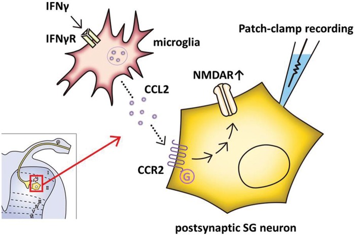
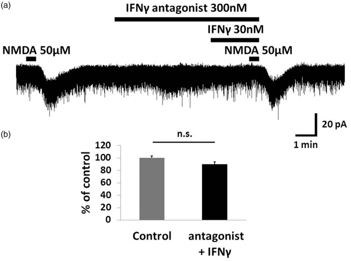
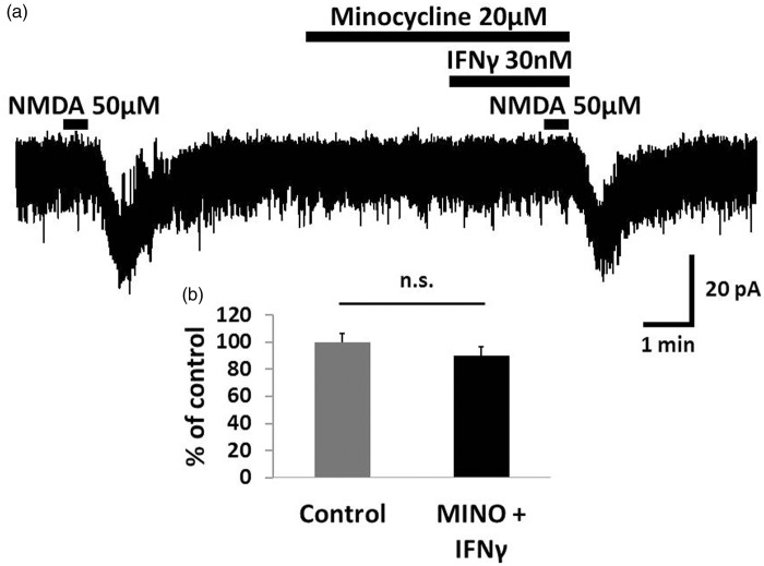
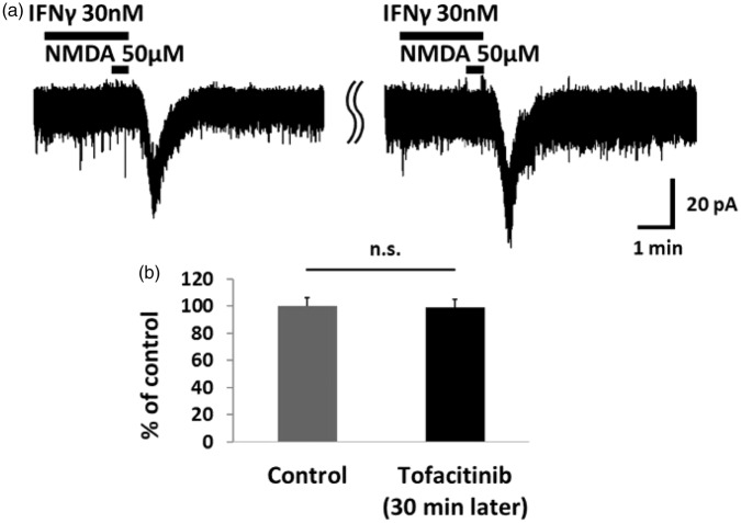
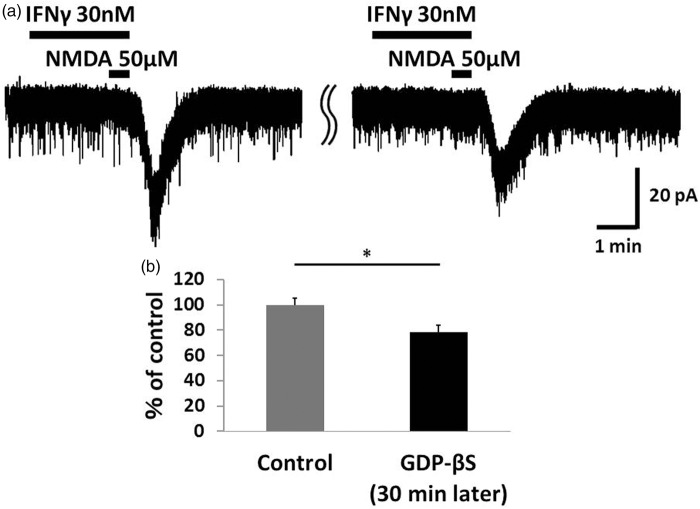
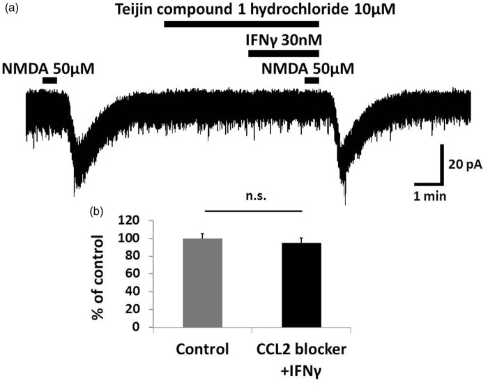
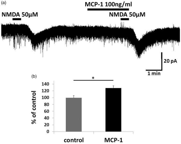
IFNγ perfusion significantly enhanced the amplitude of NMDA-induced inward currents in substantia gelatinosa neurons, but did not affect AMPA-induced currents. The facilitation of NMDA-induced current by IFNγ was inhibited by bath application of an IFNγ receptor-selective antagonist. Adding the Janus activated kinase inhibitor tofacitinib to the pipette solution did not affect the IFNγ-induced facilitation of NMDA-induced currents. However, the facilitatory effect of IFNγ on NMDA-induced currents was inhibited by perfusion of the microglial inhibitor minocycline. These results suggest that IFNγ binds the microglial IFNγ receptor and enhances NMDA receptor activity in substantia gelatinosa neurons. Next, to identify the effector of signal transmission from microglia to dorsal horn neurons, we added an inhibitor of G proteins, GDP-β-S, to the pipette solution. In a GDP-β-S-containing pipette solution, IFNγ-induced potentiation of the NMDA current was significantly suppressed after 30 min. In addition, IFNγ-induced potentiation of NMDA currents was blocked by application of a selective antagonist of CCR2, and its ligand CCL2 increased NMDA-induced currents.
Our findings suggest that IFNγ enhance the amplitude of NMDA-induced inward currents in substantia gelatinosa neurons via microglial IFNγ receptors and CCL2/CCR2 signaling. This mechanism might be partially responsible for the development of persistent neuropathic pain.
胶质细胞与神经元的相互作用在神经性疼痛的发展中起重要作用。促炎细胞因子γ干扰素(IFNγ)在外周神经损伤后脊髓背角的表达上调,鞘内注射IFNγ可诱导大鼠出现机械性异常性疼痛。越来越多的证据表明,IFNγ可能参与神经性疼痛的机制,但其对脊髓背角的影响尚不清楚。我们进行了盲法全细胞膜片钳记录,以研究IFNγ对成年雄性大鼠脊髓切片胶状质神经元中突触后谷氨酸诱导电流的影响。
IFNγ灌注显著增强了胶状质神经元中NMDA诱导的内向电流幅度,但不影响AMPA诱导的电流。IFNγ受体选择性拮抗剂的浴用抑制了IFNγ对NMDA诱导电流的促进作用。将Janus激活激酶抑制剂托法替布添加到移液管溶液中并不影响IFNγ诱导的NMDA诱导电流的促进作用。然而,小胶质细胞抑制剂米诺环素的灌注抑制了IFNγ对NMDA诱导电流的促进作用。这些结果表明,IFNγ与小胶质细胞IFNγ受体结合并增强胶状质神经元中的NMDA受体活性。接下来,为了确定从小胶质细胞到背角神经元的信号传递效应器,我们将G蛋白抑制剂GDP-β-S添加到移液管溶液中。在含有GDP-β-S的移液管溶液中,30分钟后IFNγ诱导的NMDA电流增强被显著抑制。此外,应用CCR2选择性拮抗剂可阻断IFNγ诱导的NMDA电流增强,其配体CCL2可增加NMDA诱导的电流。
我们的研究结果表明,IFNγ通过小胶质细胞IFNγ受体和CCL2/CCR2信号增强胶状质神经元中NMDA诱导的内向电流幅度。这一机制可能部分导致持续性神经性疼痛的发生。