Minicucci Marcos, Oliveira Fernando, Santos Priscila, Polegato Bertha, Roscani Meliza, Fernandes Ana Angelica, Lustosa Beatriz, Paiva Sergio, Zornoff Leonardo, Azevedo Paula
Faculdade de Medicina de Botucatu, Universidade Estadual Paulista, São Paulo, SP, Brazil.
Arq Bras Cardiol. 2016 May;106(5):396-403. doi: 10.5935/abc.20160057. Epub 2016 Apr 19.
Tobacco smoke exposure is an important risk factor for cardiac remodeling. Under this condition, inflammation, oxidative stress, energy metabolism abnormalities, apoptosis, and hypertrophy are present. Pentoxifylline has anti‑inflammatory, anti-apoptotic, anti-thrombotic and anti-proliferative properties.
The present study tested the hypothesis that pentoxifylline would attenuate cardiac remodeling induced by smoking.
Wistar rats were distributed in four groups: Control (C), Pentoxifylline (PX), Tobacco Smoke (TS), and PX-TS. After two months, echocardiography, invasive blood pressure measurement, biochemical, and histological studies were performed. The groups were compared by two-way ANOVA with a significance level of 5%.
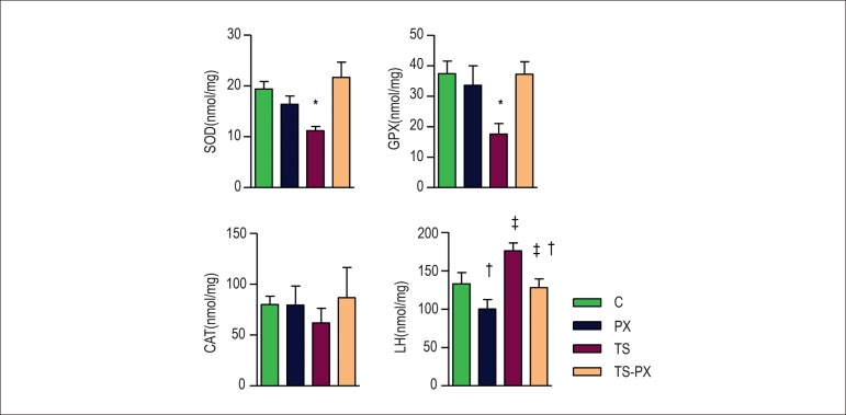
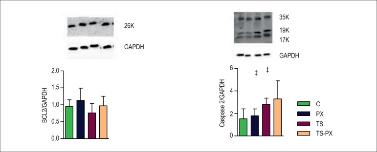
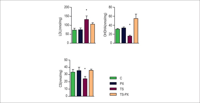
TS increased left atrium diameter and area, which was attenuated by PX. In the isolated heart study, TS lowered the positive derivate (+dp/dt), and this was attenuated by PX. The antioxidants enzyme superoxide dismutase and glutathione peroxidase were decreased in the TS group; PX recovered these activities. TS increased lactate dehydrogenase (LDH) and decreased 3-hydroxyacyl Coenzyme A dehydrogenases (OH-DHA) and citrate synthase (CS). PX attenuated LDH, 3-OH-DHA and CS alterations in TS-PX group. TS increased IL-10, ICAM-1, and caspase-3. PX did not influence these variables.
TS induced cardiac remodeling, associated with increased inflammation, oxidative stress, apoptosis, and changed energy metabolism. PX attenuated cardiac remodeling by reducing oxidative stress and improving cardiac bioenergetics, but did not act upon cardiac cytokines and apoptosis.
接触烟草烟雾是心脏重塑的一个重要风险因素。在这种情况下,会出现炎症、氧化应激、能量代谢异常、细胞凋亡和肥大。己酮可可碱具有抗炎、抗凋亡、抗血栓形成和抗增殖特性。
本研究检验了己酮可可碱可减轻吸烟诱导的心脏重塑这一假设。
将Wistar大鼠分为四组:对照组(C)、己酮可可碱组(PX)、烟草烟雾组(TS)和PX - TS组。两个月后,进行超声心动图、有创血压测量、生化和组织学研究。采用双向方差分析对各组进行比较,显著性水平为5%。
TS增加了左心房直径和面积,而PX可减轻这种增加。在离体心脏研究中,TS降低了正导数(+dp/dt),而PX可减轻这种降低。TS组抗氧化酶超氧化物歧化酶和谷胱甘肽过氧化物酶减少;PX恢复了这些活性。TS增加了乳酸脱氢酶(LDH),降低了3 - 羟基酰基辅酶A脱氢酶(OH - DHA)和柠檬酸合酶(CS)。PX减轻了TS - PX组中LDH、3 - OH - DHA和CS的改变。TS增加了白细胞介素 - 10、细胞间黏附分子 - 1和半胱天冬酶 - 3。PX对这些变量没有影响。
TS诱导心脏重塑,伴有炎症增加、氧化应激、细胞凋亡和能量代谢改变。PX通过降低氧化应激和改善心脏生物能量学减轻了心脏重塑,但对心脏细胞因子和细胞凋亡没有作用。