• 文献检索
  • 文档翻译
  • 深度研究
  • 学术资讯
  • Suppr Zotero 插件Zotero 插件
  • 邀请有礼
  • 套餐&价格
  • 历史记录
应用&插件
Suppr Zotero 插件Zotero 插件浏览器插件Mac 客户端Windows 客户端微信小程序
定价
高级版会员购买积分包购买API积分包
服务
文献检索文档翻译深度研究API 文档MCP 服务
关于我们
关于 Suppr公司介绍联系我们用户协议隐私条款
关注我们

Suppr 超能文献

核心技术专利:CN118964589B侵权必究
粤ICP备2023148730 号-1Suppr @ 2026

文献检索

告别复杂PubMed语法,用中文像聊天一样搜索,搜遍4000万医学文献。AI智能推荐,让科研检索更轻松。

立即免费搜索

文件翻译

保留排版,准确专业,支持PDF/Word/PPT等文件格式,支持 12+语言互译。

免费翻译文档

深度研究

AI帮你快速写综述,25分钟生成高质量综述,智能提取关键信息,辅助科研写作。

立即免费体验

cJun氨基末端激酶抑制剂对视网膜神经节细胞的体外和体内神经保护作用。

In vitro and in vivo neuroprotective effects of cJun N-terminal kinase inhibitors on retinal ganglion cells.

作者信息

Kim Byung-Jin, Silverman Sean M, Liu Yang, Wordinger Robert J, Pang Iok-Hou, Clark Abbot F

机构信息

North Texas Eye Research Institute, University of North Texas Health Science Center, 3500 Camp Bowie Boulevard, Fort Worth, TX, 76109, USA.

Department of Pharmaceutical Sciences, University of North Texas Health Science Center, Fort Worth, TX, 76107, USA.

出版信息

Mol Neurodegener. 2016 Apr 21;11:30. doi: 10.1186/s13024-016-0093-4.

DOI:10.1186/s13024-016-0093-4
PMID:27098079
原文链接:https://pmc.ncbi.nlm.nih.gov/articles/PMC4839164/
Abstract

BACKGROUND

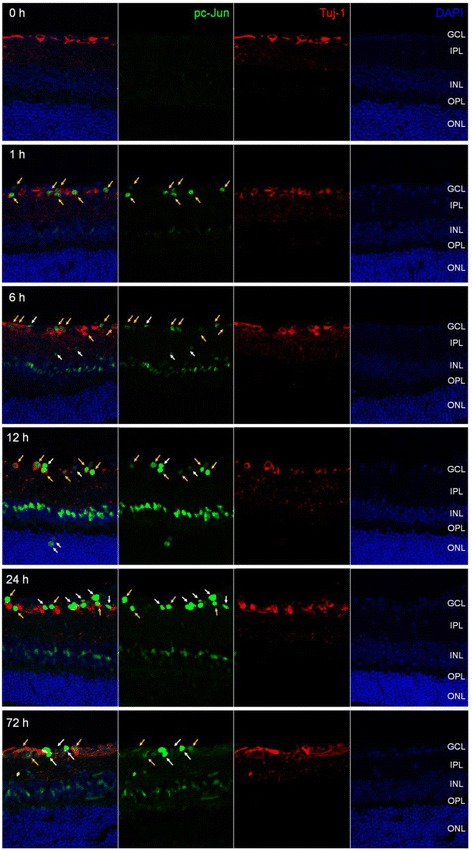
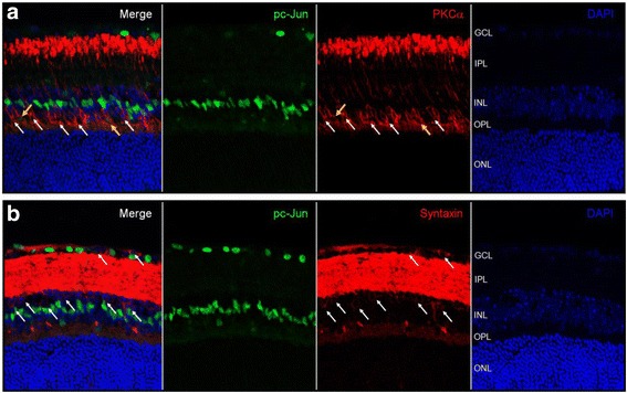
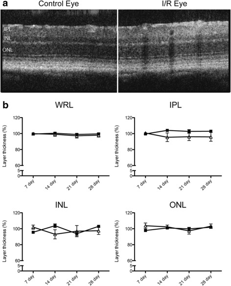
The c-Jun N-terminal kinase (JNK) signaling pathway plays an important role in neuronal pathophysiology. Using JNK inhibitors, we examined involvement of the JNK pathway in cultured rat retinal ganglion cell (RGC) death and in mouse retinal ischemia/reperfusion (I/R) injury of the visual axis. The in vitro effects of JNK inhibitors were evaluated in cultured adult rat retinal cells enriched in RGCs. Retinal I/R was induced in C57BL/6J mice through elevation of intraocular pressure to 120 mmHg for 60 min followed by reperfusion. SP600125 was administered intraperitoneally once daily for 28 days. Phosphorylation of JNK and c-Jun in the retina was examined by immunoblotting and immunohistochemistry. The thickness of retinal layers and cell numbers in the ganglion cell layer (GCL) were examined using H&E stained retinal cross sections and spectral domain optical coherence tomography (SD-OCT). Retinal function was measured by scotopic flash electroretinography (ERG). Volumetric measurement of the superior colliculus (SC) as well as VGLUT2 and PSD95 expression were studied.

RESULTS

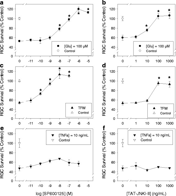
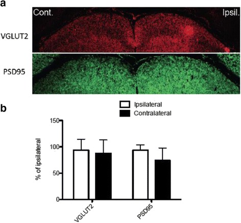
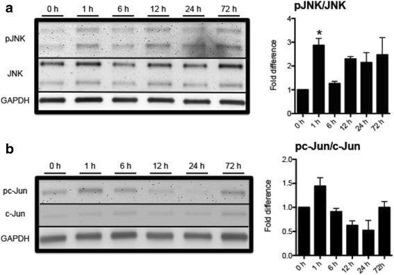
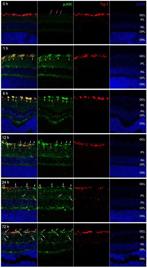
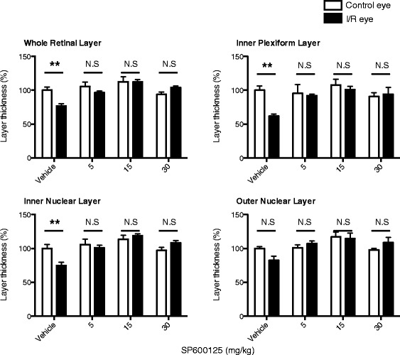
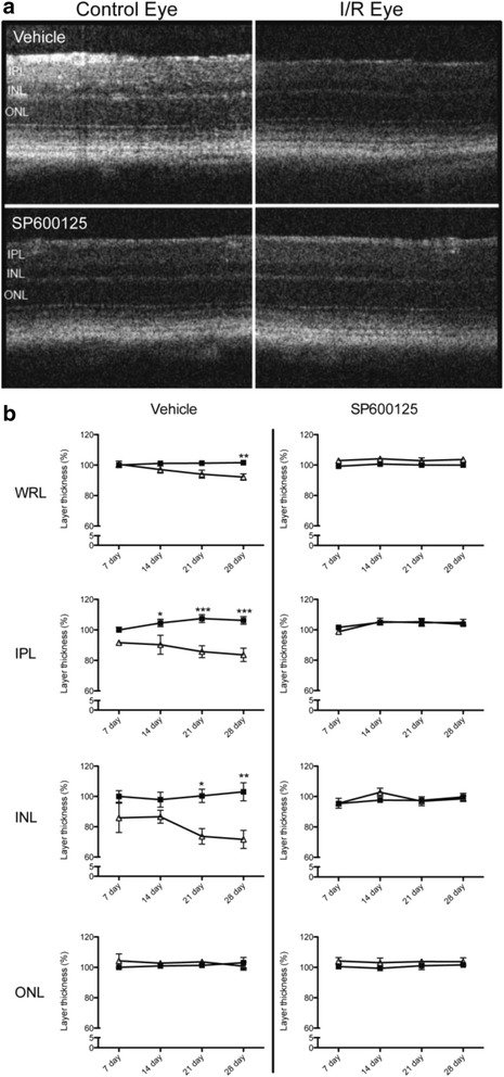
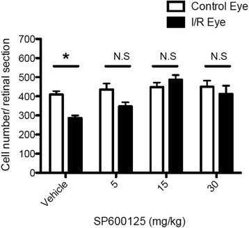
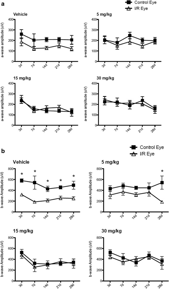
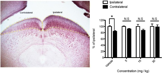
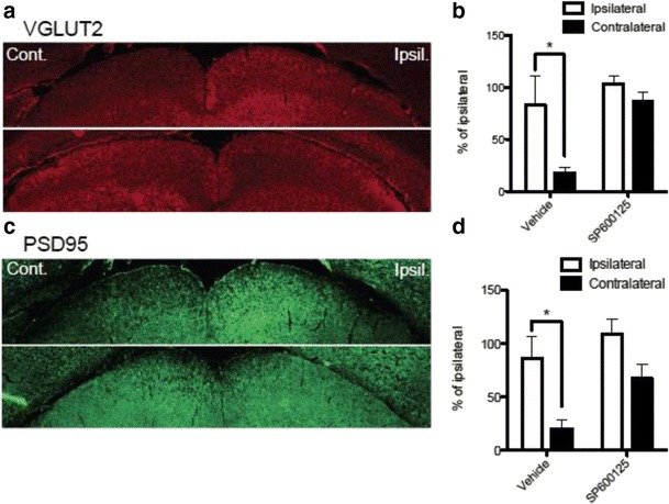
JNK inhibitors SP600125 and TAT-JNK-III, dose-dependently and significantly (p < 0.05) protected against glutamate excitotoxicity and trophic factor withdrawal induced RGC death in culture. In the I/R model, phosphorylation of JNK (pJNK) in the retina was significantly (p < 0.05) increased after injury. I/R injury significantly (p < 0.05) decreased the thickness of retinal layers, including the whole retina, inner plexiform layer, and inner nuclear layer and cell numbers in the GCL. Administration of SP600125 for 28 days protected against all these degenerative morphological changes (p < 0.05). In addition, SP600125 significantly (p < 0.05) protected against I/R-induced reduction in scotopic ERG b-wave amplitude at 3, 7, 14, 21 and 28 days after injury. SP600125 also protected against the I/R-induced losses in volume and levels of synaptic markers in the SC. Moreover, the protective effects of SP600125 in the retina and SC were also detected even with only 7 days (Days 1-7 after I/R) of SP600125 treatment.

CONCLUSIONS

Our results demonstrate the important role the JNK pathway plays in retinal degeneration in both in vitro and in vivo models and suggest that JNK inhibitors may be a useful therapeutic strategy for neuroprotection of RGCs in the retina.

摘要

背景

c-Jun氨基末端激酶(JNK)信号通路在神经元病理生理学中起重要作用。我们使用JNK抑制剂,研究了JNK通路在培养的大鼠视网膜神经节细胞(RGC)死亡以及小鼠视轴视网膜缺血/再灌注(I/R)损伤中的作用。在富含RGC的成年大鼠视网膜细胞培养物中评估JNK抑制剂的体外作用。通过将眼压升高至120 mmHg持续60分钟,然后再灌注,在C57BL/6J小鼠中诱导视网膜I/R。每天腹腔注射SP600125一次,共28天。通过免疫印迹和免疫组织化学检测视网膜中JNK和c-Jun的磷酸化。使用苏木精和伊红(H&E)染色的视网膜横断面和光谱域光学相干断层扫描(SD-OCT)检查视网膜层的厚度和神经节细胞层(GCL)中的细胞数量。通过暗视闪光视网膜电图(ERG)测量视网膜功能。研究了上丘(SC)的体积测量以及VGLUT2和PSD95的表达。

结果

JNK抑制剂SP600125和TAT-JNK-III在体外剂量依赖性且显著地(p<0.05)保护培养的RGC免受谷氨酸兴奋性毒性和营养因子剥夺诱导的死亡。在I/R模型中,损伤后视网膜中JNK(pJNK)的磷酸化显著(p<0.05)增加。I/R损伤显著(p<0.05)降低了包括整个视网膜、内网状层和内核层在内的视网膜层厚度以及GCL中的细胞数量。给予SP600125 28天可防止所有这些退行性形态学变化(p<0.05)。此外,SP600125在损伤后第3、7、14、21和28天显著(p<0.05)保护I/R诱导的暗视ERG b波振幅降低。SP600125还可防止I/R诱导的SC中突触标记物的体积和水平损失。此外,即使仅用SP600125治疗7天(I/R后的第1-7天),也能检测到SP600125对视网膜和SC的保护作用。

结论

我们的结果证明了JNK通路在体外和体内模型的视网膜变性中所起的重要作用,并表明JNK抑制剂可能是视网膜中RGC神经保护的一种有用治疗策略。

https://cdn.ncbi.nlm.nih.gov/pmc/blobs/d58b/4839164/fd883f4d7063/13024_2016_93_Fig13_HTML.jpg
https://cdn.ncbi.nlm.nih.gov/pmc/blobs/d58b/4839164/88289b7a6a17/13024_2016_93_Fig1_HTML.jpg
https://cdn.ncbi.nlm.nih.gov/pmc/blobs/d58b/4839164/2b21a4248cfb/13024_2016_93_Fig2_HTML.jpg
https://cdn.ncbi.nlm.nih.gov/pmc/blobs/d58b/4839164/2444d8c2bc9f/13024_2016_93_Fig3_HTML.jpg
https://cdn.ncbi.nlm.nih.gov/pmc/blobs/d58b/4839164/1a9cafc5828d/13024_2016_93_Fig4_HTML.jpg
https://cdn.ncbi.nlm.nih.gov/pmc/blobs/d58b/4839164/82ed690bcb57/13024_2016_93_Fig5_HTML.jpg
https://cdn.ncbi.nlm.nih.gov/pmc/blobs/d58b/4839164/879ca0196850/13024_2016_93_Fig6_HTML.jpg
https://cdn.ncbi.nlm.nih.gov/pmc/blobs/d58b/4839164/70d500337bf6/13024_2016_93_Fig7_HTML.jpg
https://cdn.ncbi.nlm.nih.gov/pmc/blobs/d58b/4839164/ba1e441c1dd6/13024_2016_93_Fig8_HTML.jpg
https://cdn.ncbi.nlm.nih.gov/pmc/blobs/d58b/4839164/abde4199bd63/13024_2016_93_Fig9_HTML.jpg
https://cdn.ncbi.nlm.nih.gov/pmc/blobs/d58b/4839164/720580aa098f/13024_2016_93_Fig10_HTML.jpg
https://cdn.ncbi.nlm.nih.gov/pmc/blobs/d58b/4839164/a135a898a8cd/13024_2016_93_Fig11_HTML.jpg
https://cdn.ncbi.nlm.nih.gov/pmc/blobs/d58b/4839164/08f0e805e66b/13024_2016_93_Fig12_HTML.jpg
https://cdn.ncbi.nlm.nih.gov/pmc/blobs/d58b/4839164/fd883f4d7063/13024_2016_93_Fig13_HTML.jpg

相似文献

1
In vitro and in vivo neuroprotective effects of cJun N-terminal kinase inhibitors on retinal ganglion cells.cJun氨基末端激酶抑制剂对视网膜神经节细胞的体外和体内神经保护作用。
Mol Neurodegener. 2016 Apr 21;11:30. doi: 10.1186/s13024-016-0093-4.
2
Protective effect of a JNK inhibitor against retinal ganglion cell loss induced by acute moderate ocular hypertension.JNK抑制剂对急性中度高眼压诱导的视网膜神经节细胞丢失的保护作用。
Mol Vis. 2011 Apr 6;17:864-75.
3
C1q propagates microglial activation and neurodegeneration in the visual axis following retinal ischemia/reperfusion injury.C1q在视网膜缺血/再灌注损伤后,在视轴中促进小胶质细胞激活和神经退行性变。
Mol Neurodegener. 2016 Mar 24;11:24. doi: 10.1186/s13024-016-0089-0.
4
Neuroprotective effects of granulocyte colony-stimulating factor on ischemia-reperfusion injury of the retina.粒细胞集落刺激因子对视网膜缺血再灌注损伤的神经保护作用。
Ophthalmic Res. 2012;48(4):199-207. doi: 10.1159/000340059. Epub 2012 Aug 3.
5
Transforming growth-beta 1 contributes to isoflurane postconditioning against cerebral ischemia-reperfusion injury by regulating the c-Jun N-terminal kinase signaling pathway.转化生长因子-β1通过调节c-Jun氨基末端激酶信号通路,有助于异氟烷对脑缺血再灌注损伤的后处理作用。
Biomed Pharmacother. 2016 Mar;78:280-290. doi: 10.1016/j.biopha.2016.01.030. Epub 2016 Feb 4.
6
Neuroprotection against focal ischemic brain injury by inhibition of c-Jun N-terminal kinase and attenuation of the mitochondrial apoptosis-signaling pathway.通过抑制c-Jun氨基末端激酶和减弱线粒体凋亡信号通路对局灶性缺血性脑损伤的神经保护作用。
J Cereb Blood Flow Metab. 2005 Jun;25(6):694-712. doi: 10.1038/sj.jcbfm.9600062.
7
Protective effects of catalase on retinal ischemia/reperfusion injury in rats.过氧化氢酶对大鼠视网膜缺血/再灌注损伤的保护作用。
Exp Eye Res. 2011 Nov;93(5):599-606. doi: 10.1016/j.exer.2011.07.007. Epub 2011 Aug 2.
8
JNK Inhibition Reduced Retinal Ganglion Cell Death after Ischemia/Reperfusion In Vivo and after Hypoxia In Vitro.JNK抑制可减少体内缺血/再灌注后及体外缺氧后的视网膜神经节细胞死亡。
Adv Exp Med Biol. 2016;854:677-83. doi: 10.1007/978-3-319-17121-0_90.
9
Role of the c-Jun N-terminal kinase pathway in retinal excitotoxicity, and neuroprotection by its inhibition.c-Jun N-端激酶通路在视网膜兴奋性毒性中的作用及其抑制的神经保护作用。
J Neurochem. 2010 Jun;113(5):1307-18. doi: 10.1111/j.1471-4159.2010.06705.x. Epub 2010 Mar 25.
10
SP600125, a selective JNK inhibitor, protects ischemic renal injury via suppressing the extrinsic pathways of apoptosis.SP600125是一种选择性JNK抑制剂,通过抑制细胞凋亡的外源性途径来保护缺血性肾损伤。
Life Sci. 2007 May 8;80(22):2067-75. doi: 10.1016/j.lfs.2007.03.010. Epub 2007 Mar 27.

引用本文的文献

1
Modulation of Oxidative Stress in Diabetic Retinopathy: Therapeutic Role of Natural Polyphenols.糖尿病视网膜病变中氧化应激的调节:天然多酚的治疗作用
Antioxidants (Basel). 2025 Jul 17;14(7):875. doi: 10.3390/antiox14070875.
2
Enhancing radiosensitivity of osteosarcoma by ITGB3 knockdown: a mechanism linked to enhanced osteogenic differentiation status through JNK/c-JUN/RUNX2 pathway activation.通过敲低整合素β3(ITGB3)增强骨肉瘤的放射敏感性:一种通过JNK/c-JUN/RUNX2途径激活与增强成骨分化状态相关的机制。
J Exp Clin Cancer Res. 2025 May 24;44(1):159. doi: 10.1186/s13046-025-03417-4.
3
The molecular mechanisms underlying retinal ganglion cell apoptosis and optic nerve regeneration in glaucoma (Review).

本文引用的文献

1
Inhibition of thioredoxin-1 with siRNA exacerbates apoptosis by activating the ASK1-JNK/p38 pathway in brain of a stroke model rats.用小干扰RNA抑制硫氧还蛋白-1可通过激活中风模型大鼠脑中的ASK1-JNK/p38信号通路加剧细胞凋亡。
Brain Res. 2015 Mar 2;1599:20-31. doi: 10.1016/j.brainres.2014.12.033. Epub 2014 Dec 22.
2
Chitosan oligosaccharides prevented retinal ischemia and reperfusion injury via reduced oxidative stress and inflammation in rats.壳寡糖通过减轻大鼠的氧化应激和炎症反应预防视网膜缺血再灌注损伤。
Exp Eye Res. 2015 Jan;130:38-50. doi: 10.1016/j.exer.2014.12.001. Epub 2014 Dec 2.
3
A combination of four active compounds alleviates cerebral ischemia-reperfusion injury in correlation with inhibition of autophagy and modulation of AMPK/mTOR and JNK pathways.
青光眼视网膜神经节细胞凋亡及视神经再生的分子机制(综述)
Int J Mol Med. 2025 Apr;55(4). doi: 10.3892/ijmm.2025.5504. Epub 2025 Feb 14.
4
The Impact of Aging on Ocular Diseases: Unveiling Complex Interactions.衰老对眼部疾病的影响:揭示复杂的相互作用
Aging Dis. 2024 Oct 17. doi: 10.14336/AD.2024.0850.
5
Diabetic Retinopathy: New Treatment Approaches Targeting Redox and Immune Mechanisms.糖尿病视网膜病变:针对氧化还原和免疫机制的新治疗方法
Antioxidants (Basel). 2024 May 12;13(5):594. doi: 10.3390/antiox13050594.
6
Modeling of Retina and Optic Nerve Ischemia-Reperfusion Injury through Hypoxia-Reoxygenation in Human Induced Pluripotent Stem Cell-Derived Retinal Ganglion Cells.通过人诱导多能干细胞衍生的视网膜神经节细胞中的缺氧-复氧模拟视网膜和视神经缺血再灌注损伤。
Cells. 2024 Jan 11;13(2):130. doi: 10.3390/cells13020130.
7
Oxidative stress in the eye and its role in the pathophysiology of ocular diseases.眼睛中的氧化应激及其在眼部疾病病理生理学中的作用。
Redox Biol. 2023 Dec;68:102967. doi: 10.1016/j.redox.2023.102967. Epub 2023 Nov 18.
8
Keratin8 Deficiency Aggravates Retinal Ganglion Cell Damage Under Acute Ocular Hypertension.角蛋白 8 缺乏症在急性眼高压下加重视网膜神经节细胞损伤。
Invest Ophthalmol Vis Sci. 2023 Sep 1;64(12):1. doi: 10.1167/iovs.64.12.1.
9
Reactive Bergmann glia play a central role in spinocerebellar ataxia inflammation via the JNK pathway.反应性 Bergmann 胶质细胞通过 JNK 通路在脊髓小脑共济失调炎症中发挥核心作用。
J Neuroinflammation. 2023 May 26;20(1):126. doi: 10.1186/s12974-023-02801-1.
10
Implications of Diabetes-Induced Altered Metabolites on Retinal Neurodegeneration.糖尿病诱导的代谢物改变对视网膜神经退行性变的影响。
Front Neurosci. 2022 Jul 13;16:938029. doi: 10.3389/fnins.2022.938029. eCollection 2022.
四种活性化合物的组合通过抑制自噬以及调节AMPK/mTOR和JNK信号通路来减轻脑缺血再灌注损伤。
J Neurosci Res. 2014 Oct;92(10):1295-306. doi: 10.1002/jnr.23400. Epub 2014 May 7.
4
Loss of inner retinal neurons after retinal ischemia in rats.大鼠视网膜缺血后视网膜内层神经元的丢失。
Invest Ophthalmol Vis Sci. 2014 Apr 28;55(4):2777-87. doi: 10.1167/iovs.13-13372.
5
Transplantation with retinal progenitor cells repairs visual function in rats with retinal ischemia-reperfusion injury.视网膜祖细胞移植可修复视网膜缺血再灌注损伤大鼠的视觉功能。
Neurosci Lett. 2014 Jan 13;558:8-13. doi: 10.1016/j.neulet.2013.09.024. Epub 2013 Sep 25.
6
JNK-mediated phosphorylation of DLK suppresses its ubiquitination to promote neuronal apoptosis.JNK 介导的 DLK 磷酸化抑制其泛素化,从而促进神经元凋亡。
J Cell Biol. 2013 Sep 2;202(5):747-63. doi: 10.1083/jcb.201303066. Epub 2013 Aug 26.
7
Progressive morphological changes and impaired retinal function associated with temporal regulation of gene expression after retinal ischemia/reperfusion injury in mice.视网膜缺血/再灌注损伤后,小鼠基因表达的时间调控与进行性形态变化和视网膜功能障碍有关。
Mol Neurodegener. 2013 Jun 22;8:21. doi: 10.1186/1750-1326-8-21.
8
DLK initiates a transcriptional program that couples apoptotic and regenerative responses to axonal injury.DLK 启动了一个转录程序,将细胞凋亡和再生反应与轴突损伤联系起来。
Proc Natl Acad Sci U S A. 2013 Mar 5;110(10):4039-44. doi: 10.1073/pnas.1211074110. Epub 2013 Feb 19.
9
Functional genomic screening identifies dual leucine zipper kinase as a key mediator of retinal ganglion cell death.功能基因组筛选鉴定双亮氨酸拉链激酶为视网膜神经节细胞死亡的关键介质。
Proc Natl Acad Sci U S A. 2013 Mar 5;110(10):4045-50. doi: 10.1073/pnas.1211284110. Epub 2013 Feb 19.
10
Crocetin, a carotenoid derivative, inhibits retinal ischemic damage in mice.西红花酸,类胡萝卜素的衍生物,可抑制小鼠视网膜缺血损伤。
Eur J Pharmacol. 2013 Mar 5;703(1-3):1-10. doi: 10.1016/j.ejphar.2013.02.007. Epub 2013 Feb 18.