Park Bumhee, Palomares Jose A, Woo Mary A, Kang Daniel W, Macey Paul M, Yan-Go Frisca L, Harper Ronald M, Kumar Rajesh
Department of Anesthesiology University of California at Los Angeles Los Angeles CA 90095.
UCLA School of Nursing University of California at Los Angeles Los Angeles CA 90095.
Brain Behav. 2016 Feb 1;6(3):e00441. doi: 10.1002/brb3.441. eCollection 2016 Mar.
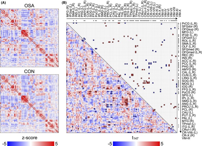
Obstructive sleep apnea (OSA) subjects show impaired autonomic, affective, executive, sensorimotor, and cognitive functions. Brain injury in OSA subjects appears in multiple sites regulating these functions, but the integrity of functional networks within the regulatory sites remains unclear. Our aim was to examine the functional interactions and the complex network organization of these interactions across the whole brain in OSA, using regional functional connectivity (FC) and brain network topological properties.
We collected resting-state functional magnetic resonance imaging (MRI) data, using a 3.0-Tesla MRI scanner, from 69 newly diagnosed, treatment-naïve, moderate-to-severe OSA (age, 48.3 ± 9.2 years; body mass index, 31 ± 6.2 kg/m(2); apnea-hypopnea index (AHI), 35.6 ± 23.3 events/h) and 82 control subjects (47.6 ± 9.1 years; body mass index, 25.1 ± 3.5 kg/m(2)). Data were analyzed to examine FC in OSA over controls as interregional correlations and brain network topological properties.
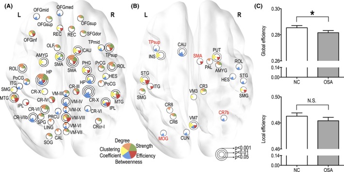
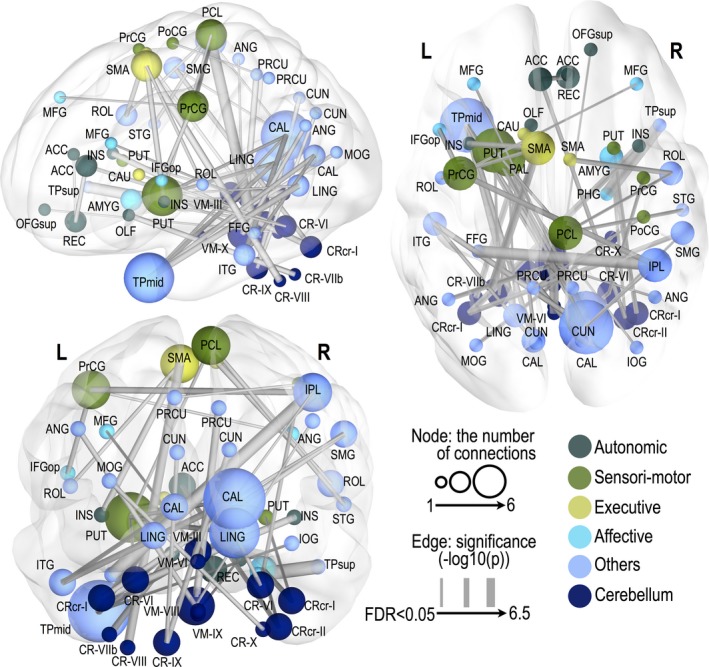
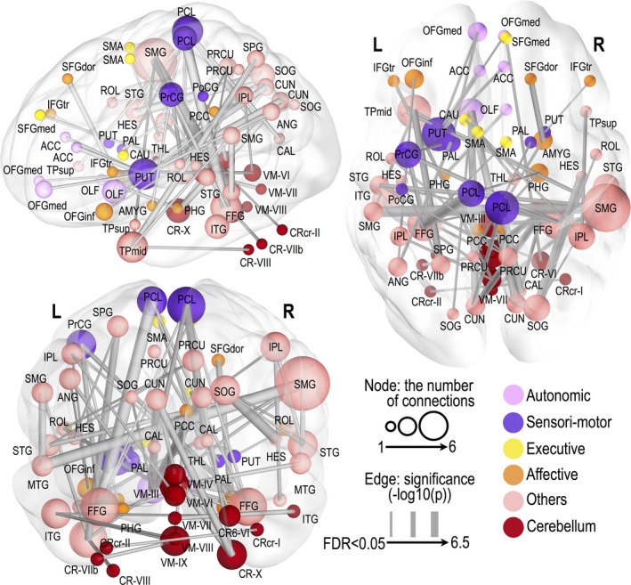
Obstructive sleep apnea subjects showed significantly altered FC in the cerebellar, frontal, parietal, temporal, occipital, limbic, and basal ganglia regions (FDR, P < 0.05). Entire functional brain networks in OSA subjects showed significantly less efficient integration, and their regional topological properties of functional integration and specialization characteristics also showed declined trends in areas showing altered FC, an outcome which would interfere with brain network organization (P < 0.05; 10,000 permutations). Brain sites with abnormal topological properties in OSA showed significant relationships with AHI scores.
Our findings suggest that the dysfunction extends to resting conditions, and the altered FC and impaired network organization may underlie the impaired responses in autonomic, cognitive, and sensorimotor functions. The outcomes likely result from the prominent structural changes in both axons and nuclear structures, which occur in the condition.
阻塞性睡眠呼吸暂停(OSA)患者表现出自主神经、情感、执行、感觉运动和认知功能受损。OSA患者的脑损伤出现在多个调节这些功能的部位,但调节部位内功能网络的完整性仍不清楚。我们的目的是使用区域功能连接(FC)和脑网络拓扑特性,研究OSA患者全脑范围内这些相互作用的功能交互和复杂网络组织。
我们使用3.0特斯拉磁共振成像(MRI)扫描仪,收集了69名新诊断的、未经治疗的中重度OSA患者(年龄48.3±9.2岁;体重指数31±6.2kg/m²;呼吸暂停低通气指数(AHI)35.6±23.3次/小时)和82名对照者(47.6±9.1岁;体重指数25.1±3.5kg/m²)的静息态功能磁共振成像数据。对数据进行分析,以检查OSA患者相对于对照者的FC,作为区域间相关性和脑网络拓扑特性。
阻塞性睡眠呼吸暂停患者在小脑、额叶、顶叶、颞叶、枕叶、边缘叶和基底神经节区域的FC有显著改变(FDR,P<0.05)。OSA患者的整个功能性脑网络显示出整合效率显著降低,其功能整合和专业化特征的区域拓扑特性在显示FC改变的区域也呈现下降趋势,这一结果会干扰脑网络组织(P<0.05;10,000次排列)。OSA患者中拓扑特性异常的脑区与AHI评分有显著关系。
我们的研究结果表明,功能障碍延伸至静息状态,FC改变和网络组织受损可能是自主神经、认知和感觉运动功能反应受损的基础。这些结果可能是由于该疾病中轴突和核结构均出现的显著结构变化所致。