Cole Mark A, Abd Jamil Amira H, Heather Lisa C, Murray Andrew J, Sutton Elizabeth R, Slingo Mary, Sebag-Montefiore Liam, Tan Suat Cheng, Aksentijević Dunja, Gildea Ottilie S, Stuckey Daniel J, Yeoh Kar Kheng, Carr Carolyn A, Evans Rhys D, Aasum Ellen, Schofield Christopher J, Ratcliffe Peter J, Neubauer Stefan, Robbins Peter A, Clarke Kieran
Department of Physiology, Anatomy and Genetics, University of Oxford, Oxford, United Kingdom;
Division of Cardiovascular Medicine, Radcliffe Department of Medicine, Wellcome Trust Centre for Human Genetics, University of Oxford, Oxford, United Kingdom;
FASEB J. 2016 Aug;30(8):2684-97. doi: 10.1096/fj.201500094R. Epub 2016 Apr 21.
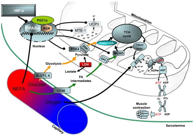
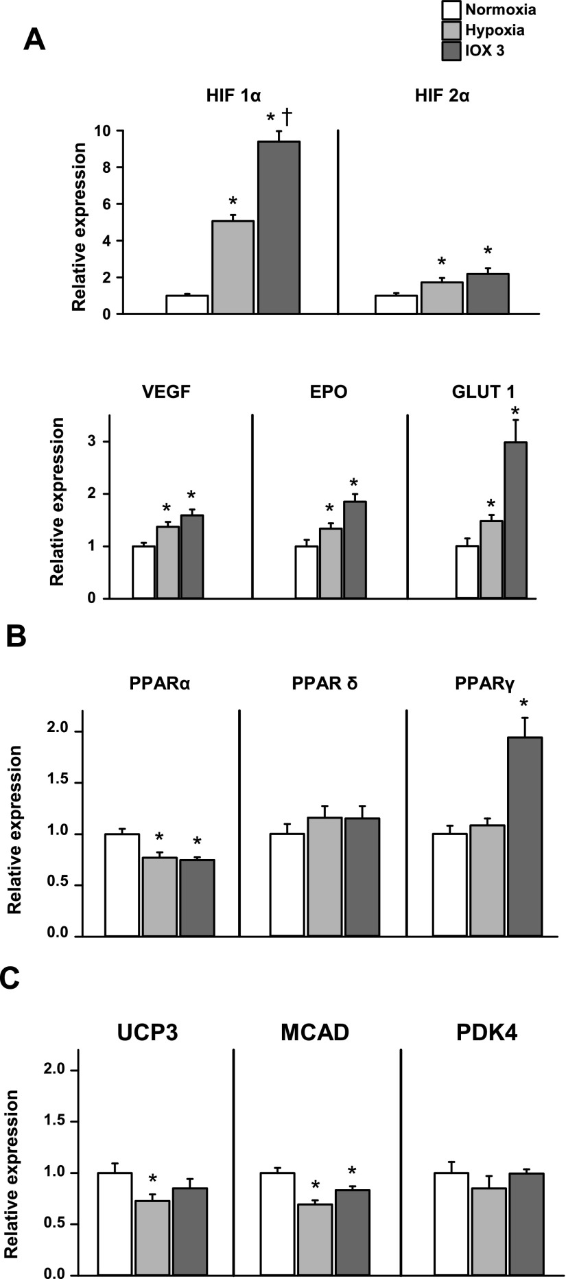
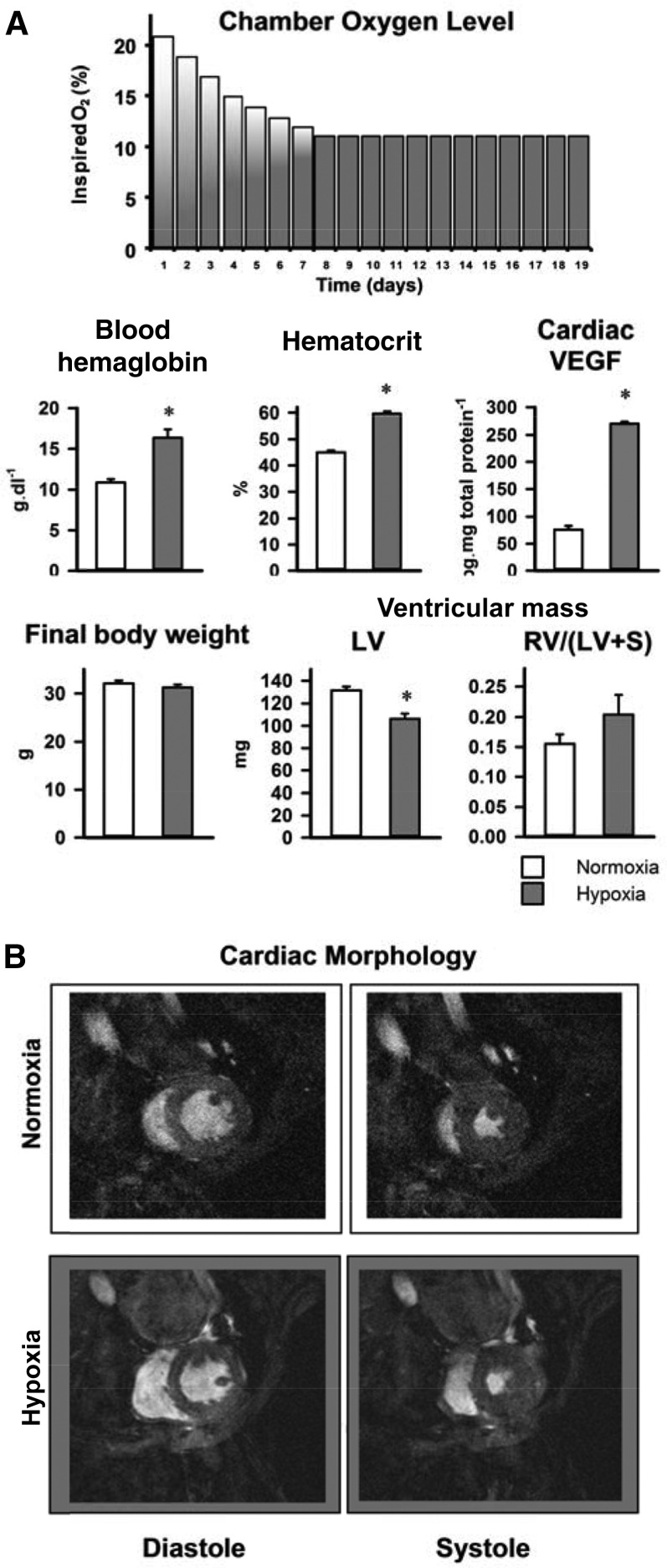
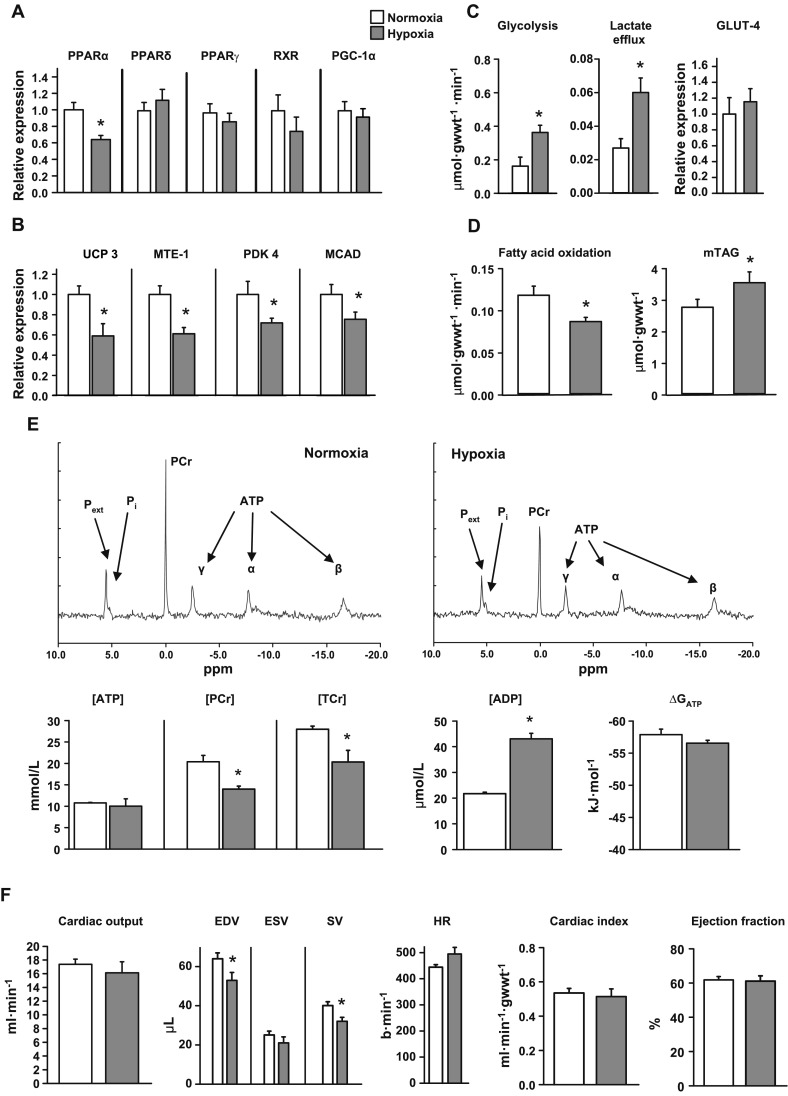
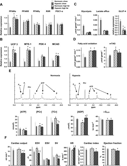
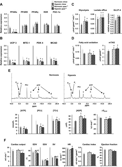
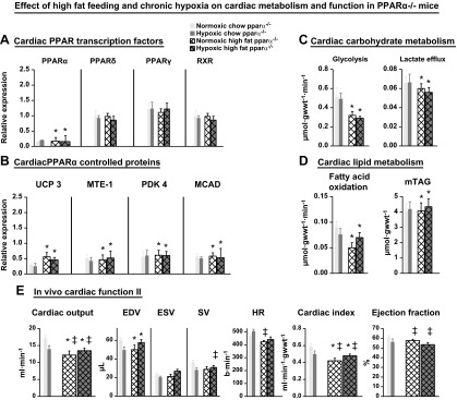
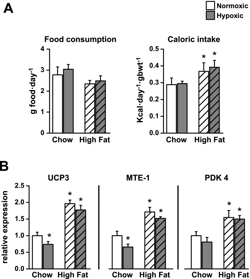
The role of peroxisome proliferator-activated receptor α (PPARα)-mediated metabolic remodeling in cardiac adaptation to hypoxia has yet to be defined. Here, mice were housed in hypoxia for 3 wk before in vivo contractile function was measured using cine MRI. In isolated, perfused hearts, energetics were measured using (31)P magnetic resonance spectroscopy (MRS), and glycolysis and fatty acid oxidation were measured using [(3)H] labeling. Compared with a normoxic, chow-fed control mouse heart, hypoxia decreased PPARα expression, fatty acid oxidation, and mitochondrial uncoupling protein 3 (UCP3) levels, while increasing glycolysis, all of which served to maintain normal ATP concentrations ([ATP]) and thereby, ejection fractions. A high-fat diet increased cardiac PPARα expression, fatty acid oxidation, and UCP3 levels with decreased glycolysis. Hypoxia was unable to alter the high PPARα expression or reverse the metabolic changes caused by the high-fat diet, with the result that [ATP] and contractile function decreased significantly. The adaptive metabolic changes caused by hypoxia in control mouse hearts were found to have occurred already in PPARα-deficient (PPARα(-/-)) mouse hearts and sustained function in hypoxia despite an inability for further metabolic remodeling. We conclude that decreased cardiac PPARα expression is essential for adaptive metabolic remodeling in hypoxia, but is prevented by dietary fat.-Cole, M. A., Abd Jamil, A. H., Heather, L. C., Murray, A. J., Sutton, E. R., Slingo, M., Sebag-Montefiore, L., Tan, S. C., Aksentijević, D., Gildea, O. S., Stuckey, D. J., Yeoh, K. K., Carr, C. A., Evans, R. D., Aasum, E., Schofield, C. J., Ratcliffe, P. J., Neubauer, S., Robbins, P. A., Clarke, K. On the pivotal role of PPARα in adaptation of the heart to hypoxia and why fat in the diet increases hypoxic injury.
过氧化物酶体增殖物激活受体α(PPARα)介导的代谢重塑在心脏适应缺氧过程中的作用尚未明确。在此,将小鼠置于缺氧环境中3周,然后使用电影磁共振成像(cine MRI)测量其体内收缩功能。在离体灌注心脏中,使用磷-31磁共振波谱(MRS)测量能量代谢,使用[氚-3]标记测量糖酵解和脂肪酸氧化。与常氧、普通饮食喂养的对照小鼠心脏相比,缺氧降低了PPARα表达、脂肪酸氧化和线粒体解偶联蛋白3(UCP3)水平,同时增加了糖酵解,所有这些都有助于维持正常的三磷酸腺苷浓度([ATP]),从而维持射血分数。高脂饮食增加了心脏PPARα表达、脂肪酸氧化和UCP3水平,同时降低了糖酵解。缺氧无法改变高脂饮食引起的高PPARα表达或逆转代谢变化,结果[ATP]和收缩功能显著下降。发现在对照小鼠心脏中由缺氧引起的适应性代谢变化在PPARα缺陷(PPARα(-/-))小鼠心脏中已经发生,并且尽管无法进行进一步的代谢重塑,但在缺氧状态下仍能维持功能。我们得出结论,心脏PPARα表达降低对于缺氧时的适应性代谢重塑至关重要,但饮食中的脂肪会阻止这种情况发生。-科尔,M.A.,阿卜杜勒·贾米尔,A.H.,希瑟,L.C.,默里,A.J.,萨顿,E.R.,斯林戈,M.,塞巴格-蒙特菲奥里,L.,谭,S.C.,阿克森蒂耶维奇,D.,吉尔迪亚,O.S.,斯塔基,D.J.,杨,K.K.,卡尔,C.A.,埃文斯,R.D.,阿苏姆,E.,斯科菲尔德,C.J.,拉特克利夫,P.J.,诺伊鲍尔,S.,罗宾斯,P.A.,克拉克,K. 关于PPARα在心脏适应缺氧中的关键作用以及饮食中的脂肪为何会增加缺氧损伤的研究。