• 文献检索
  • 文档翻译
  • 深度研究
  • 学术资讯
  • Suppr Zotero 插件Zotero 插件
  • 邀请有礼
  • 套餐&价格
  • 历史记录
应用&插件
Suppr Zotero 插件Zotero 插件浏览器插件Mac 客户端Windows 客户端微信小程序
定价
高级版会员购买积分包购买API积分包
服务
文献检索文档翻译深度研究API 文档MCP 服务
关于我们
关于 Suppr公司介绍联系我们用户协议隐私条款
关注我们

Suppr 超能文献

核心技术专利:CN118964589B侵权必究
粤ICP备2023148730 号-1Suppr @ 2026

文献检索

告别复杂PubMed语法,用中文像聊天一样搜索,搜遍4000万医学文献。AI智能推荐,让科研检索更轻松。

立即免费搜索

文件翻译

保留排版,准确专业,支持PDF/Word/PPT等文件格式,支持 12+语言互译。

免费翻译文档

深度研究

AI帮你快速写综述,25分钟生成高质量综述,智能提取关键信息,辅助科研写作。

立即免费体验

糖尿病大鼠股骨的微型计算机断层扫描:小梁骨和皮质骨微结构及血管的改变——一项可行性研究。

Microcomputed tomography of the femur of diabetic rats: alterations of trabecular and cortical bone microarchitecture and vasculature-a feasibility study.

作者信息

Zeitoun David, Caliaperoumal Guavri, Bensidhoum Morad, Constans Jean Marc, Anagnostou Fani, Bousson Valérie

机构信息

Centre hospitalier Lariboisière, Hopital Lariboisière, Service de radiologie ostéo-articulaire, 2 rue Ambroise Paré, 75010, Paris, France.

CNRS Laboratoire B2OA, Laboratoire B2OA.10, Avenue de Verdun, 75010, Paris, France.

出版信息

Eur Radiol Exp. 2019 Apr 11;3(1):17. doi: 10.1186/s41747-019-0094-5.

DOI:10.1186/s41747-019-0094-5
PMID:30972589
原文链接:https://pmc.ncbi.nlm.nih.gov/articles/PMC6458201/
Abstract

BACKGROUND

To better understand bone fragility in type 2 diabetes mellitus and define the contribution of microcomputed tomography (micro-CT) to the evaluation of bone microarchitecture and vascularisation, we conducted an in vitro preliminary study on the femur of Zucker diabetic fatty (ZDF) rats and Zucker lean (ZL) rats. We first analysed bone microarchitecture, then determined whether micro-CT allowed to explore bone vascularisation, and finally looked for a link between these parameters.

METHODS

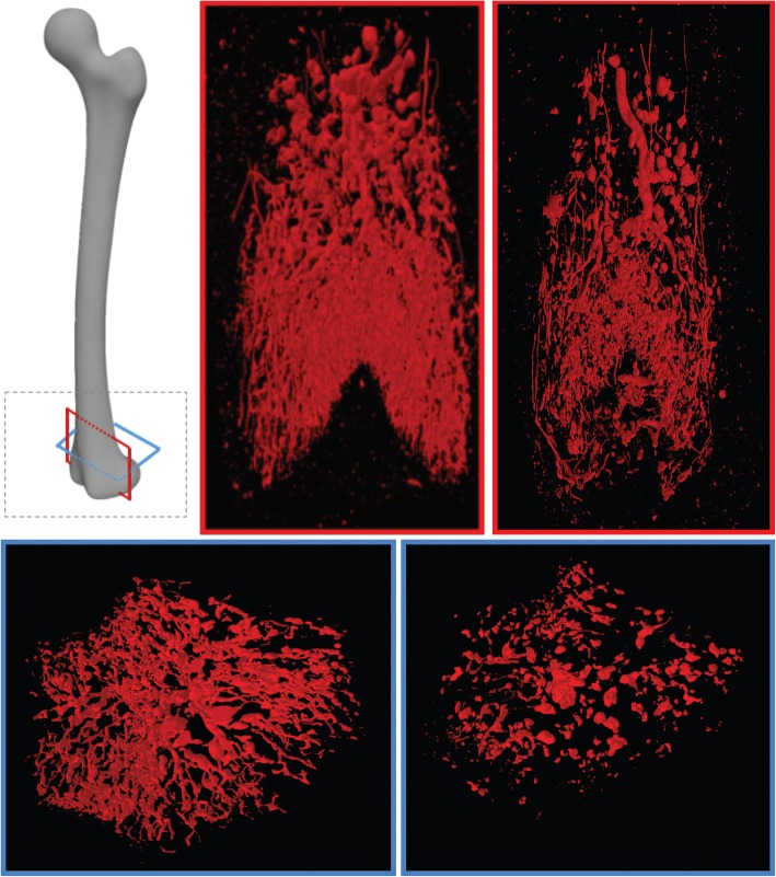
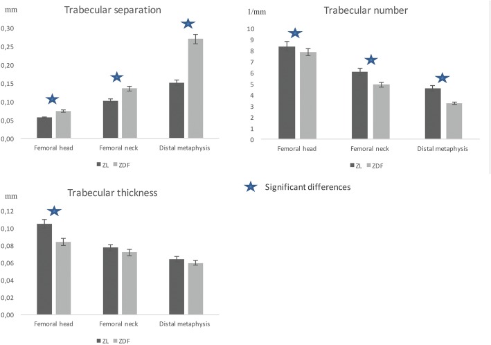
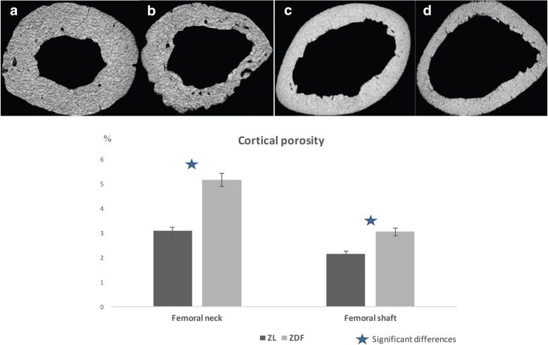
Eight ZDF and six ZL rats were examined for bone microarchitecture (group 1), and six ZDF and six ZL rats were studied for bone vascularisation after Microfil® perfusion which is a radiopaque casting agent (group 2). In group 1, we used micro-CT to examine the trabecular and cortical bone microarchitecture of the femoral head, neck, shaft, and distal metaphysis. In group 2, micro-CT was used to study the blood vessels in the head, neck, and distal metaphysis.

RESULTS

Compared to ZL rats, the ZDF rats exhibited significantly lower trabecular bone volume and number and higher trabecular separation in the three locations (p = 0.02, p = 0.02, p = 0.003). Cortical porosity was significantly higher in the ZDF rats at the neck and shaft (p = 0.001 and p = 0.005). We observed a dramatically poorer bone vascularisation in the femur of ZDF rats, especially in distal metaphysis (p < 0.047).

CONCLUSIONS

Micro-CT demonstrated not only significant alterations in the bone microarchitecture of the femurs of ZDF rats, but also significant alterations in bone vascularisation. Further studies are required to demonstrate the causal link between poor vascularisation and impaired bone architecture.

摘要

背景

为了更好地理解2型糖尿病中的骨脆性,并确定微计算机断层扫描(micro-CT)对骨微结构和血管化评估的贡献,我们对Zucker糖尿病肥胖(ZDF)大鼠和Zucker瘦素(ZL)大鼠的股骨进行了一项体外初步研究。我们首先分析骨微结构,然后确定micro-CT是否能够探究骨血管化,最后寻找这些参数之间的联系。

方法

对8只ZDF大鼠和6只ZL大鼠进行骨微结构检查(第1组),对6只ZDF大鼠和6只ZL大鼠在使用不透射线的铸型剂Microfil®灌注后进行骨血管化研究(第2组)。在第1组中,我们使用micro-CT检查股骨头、颈、骨干和干骺端的小梁骨和皮质骨微结构。在第2组中,使用micro-CT研究头、颈和干骺端的血管。

结果

与ZL大鼠相比,ZDF大鼠在三个部位的小梁骨体积和数量显著降低,小梁间距更高(p = 0.02,p = 0.02,p = 0.003)。ZDF大鼠在颈部和骨干的皮质孔隙率显著更高(p = 0.001和p = 0.005)。我们观察到ZDF大鼠股骨的骨血管化明显较差,尤其是在干骺端(p < 0.047)。

结论

Micro-CT不仅显示ZDF大鼠股骨的骨微结构有显著改变,而且骨血管化也有显著改变。需要进一步研究来证明血管化不良与骨结构受损之间的因果关系。

https://cdn.ncbi.nlm.nih.gov/pmc/blobs/e3fc/6458201/a18c2118c303/41747_2019_94_Fig4_HTML.jpg
https://cdn.ncbi.nlm.nih.gov/pmc/blobs/e3fc/6458201/9e5b549cd589/41747_2019_94_Fig1_HTML.jpg
https://cdn.ncbi.nlm.nih.gov/pmc/blobs/e3fc/6458201/ba3add67c1c0/41747_2019_94_Fig2_HTML.jpg
https://cdn.ncbi.nlm.nih.gov/pmc/blobs/e3fc/6458201/246f2f407041/41747_2019_94_Fig3_HTML.jpg
https://cdn.ncbi.nlm.nih.gov/pmc/blobs/e3fc/6458201/a18c2118c303/41747_2019_94_Fig4_HTML.jpg

相似文献

1
Microcomputed tomography of the femur of diabetic rats: alterations of trabecular and cortical bone microarchitecture and vasculature-a feasibility study.糖尿病大鼠股骨的微型计算机断层扫描:小梁骨和皮质骨微结构及血管的改变——一项可行性研究。
Eur Radiol Exp. 2019 Apr 11;3(1):17. doi: 10.1186/s41747-019-0094-5.
2
Cortical Bone Morphological and Trabecular Bone Microarchitectural Changes in the Mandible and Femoral Neck of Ovariectomized Rats.去卵巢大鼠下颌骨和股骨颈皮质骨形态及小梁骨微结构变化
PLoS One. 2016 Apr 29;11(4):e0154367. doi: 10.1371/journal.pone.0154367. eCollection 2016.
3
Sclerostin does not play a major role in the pathogenesis of skeletal complications in type 2 diabetes mellitus.硬化蛋白在2型糖尿病骨骼并发症的发病机制中不起主要作用。
Osteoporos Int. 2017 Jan;28(1):309-320. doi: 10.1007/s00198-016-3718-0. Epub 2016 Jul 28.
4
Abaloparatide improves cortical geometry and trabecular microarchitecture and increases vertebral and femoral neck strength in a rat model of male osteoporosis.阿巴洛肽可改善雄性骨质疏松症大鼠模型的皮质几何形状和小梁微结构,并增加椎体和股骨颈的强度。
Bone. 2019 Jul;124:148-157. doi: 10.1016/j.bone.2019.04.025. Epub 2019 Apr 30.
5
A comparison of micro-CT and dental CT in assessing cortical bone morphology and trabecular bone microarchitecture.微计算机断层扫描与牙科计算机断层扫描在评估皮质骨形态和小梁骨微结构方面的比较。
PLoS One. 2014 Sep 16;9(9):e107545. doi: 10.1371/journal.pone.0107545. eCollection 2014.
6
Type 2 diabetes impairs angiogenesis and osteogenesis in calvarial defects: MicroCT study in ZDF rats.2 型糖尿病损害颅骨缺损中的血管生成和成骨:ZDF 大鼠的 microCT 研究。
Bone. 2018 Jul;112:161-172. doi: 10.1016/j.bone.2018.04.009. Epub 2018 Apr 25.
7
Effects of insulin therapy on porosity, non-enzymatic glycation and mechanical competence in the bone of rats with type 2 diabetes mellitus.胰岛素治疗对2型糖尿病大鼠骨骼孔隙率、非酶糖基化及力学性能的影响。
Bone. 2016 Oct;91:186-93. doi: 10.1016/j.bone.2016.08.003. Epub 2016 Aug 4.
8
The trabecular bone score: Relationships with trabecular and cortical microarchitecture measured by HR-pQCT and histomorphometry in patients with chronic kidney disease.骨小梁评分:与 HR-pQCT 和组织形态计量学测量的慢性肾脏病患者的骨小梁和皮质微结构的关系。
Bone. 2018 Nov;116:215-220. doi: 10.1016/j.bone.2018.08.006. Epub 2018 Aug 8.
9
Cortical and trabecular bone microarchitecture as an independent predictor of incident fracture risk in older women and men in the Bone Microarchitecture International Consortium (BoMIC): a prospective study.皮质骨和小梁骨微结构作为骨微结构国际联合会(BoMIC)中老年男女新发骨折风险的独立预测因子:一项前瞻性研究。
Lancet Diabetes Endocrinol. 2019 Jan;7(1):34-43. doi: 10.1016/S2213-8587(18)30308-5. Epub 2018 Nov 28.
10
Deterioration of Cortical Bone Microarchitecture: Critical Component of Renal Osteodystrophy Evaluation.皮质骨微观结构恶化:肾性骨营养不良评估的关键组成部分。
Am J Nephrol. 2018;47(6):376-384. doi: 10.1159/000489671. Epub 2018 May 23.

引用本文的文献

1
A Review of Animal Models for Studying Bone Health in Type-2 Diabetes Mellitus (T2DM) and Obesity.探讨 2 型糖尿病(T2DM)和肥胖症中骨健康的动物模型综述。
Int J Mol Sci. 2024 Aug 29;25(17):9399. doi: 10.3390/ijms25179399.
2
Strategies for Improving Impaired Osseointegration in Compromised Animal Models.改善受损动物模型中骨整合受损的策略。
J Dent Res. 2024 May;103(5):467-476. doi: 10.1177/00220345241231777. Epub 2024 Apr 15.
3
Reduced Bone Regeneration in Rats With Type 2 Diabetes Mellitus as a Result of Impaired Stromal Cell and Osteoblast Function-A Computer Modeling Study.

本文引用的文献

1
Effect of type 2 diabetes-related non-enzymatic glycation on bone biomechanical properties.2型糖尿病相关的非酶糖基化对骨生物力学性能的影响。
Bone. 2016 Jan;82:21-7. doi: 10.1016/j.bone.2015.07.028. Epub 2015 Jul 23.
2
Determinants of bone strength and quality in diabetes mellitus in humans.人类糖尿病中骨强度和质量的决定因素。
Bone. 2016 Jan;82:28-34. doi: 10.1016/j.bone.2015.07.027. Epub 2015 Jul 26.
3
Epidemiology of fractures in type 2 diabetes.2型糖尿病患者骨折的流行病学
2型糖尿病大鼠骨再生减少是由于基质细胞和成骨细胞功能受损——一项计算机建模研究
JBMR Plus. 2023 Oct 2;7(11):e10809. doi: 10.1002/jbm4.10809. eCollection 2023 Nov.
4
Unlocking the Full Potential of SGLT2 Inhibitors: Expanding Applications beyond Glycemic Control.解锁 SGLT2 抑制剂的全部潜力:超越血糖控制的应用拓展。
Int J Mol Sci. 2023 Mar 23;24(7):6039. doi: 10.3390/ijms24076039.
5
Diabetes mellitus impairs bone regeneration and biomechanics.糖尿病会损害骨再生和生物力学。
J Orthop Surg Res. 2023 Mar 6;18(1):169. doi: 10.1186/s13018-023-03644-5.
6
Temporal and spatial changes in bone mineral content and mechanical properties during breast-cancer bone metastases.乳腺癌骨转移过程中骨矿物质含量和力学性能的时空变化。
Bone Rep. 2022 Jun 12;17:101597. doi: 10.1016/j.bonr.2022.101597. eCollection 2022 Dec.
7
Investigation of the effects of eugenol and quercetin on bone loss in STZ-NA induced diabetic rats utilizing micro CT.利用微型计算机断层扫描研究丁香酚和槲皮素对链脲佐菌素-烟酰胺诱导的糖尿病大鼠骨质流失的影响。
J Diabetes Metab Disord. 2022 Mar 12;21(1):637-646. doi: 10.1007/s40200-022-01026-y. eCollection 2022 Jun.
8
Comparison of Quantitative Computed Tomography and Dual X-Ray Absorptiometry: Osteoporosis Detection Rates in Diabetic Patients.定量计算机断层扫描与双能X线吸收法的比较:糖尿病患者的骨质疏松症检出率
Cureus. 2022 Mar 13;14(3):e23131. doi: 10.7759/cureus.23131. eCollection 2022 Mar.
9
Prediction of Femoral Strength Based on Bone Density and Biochemical Markers in Elderly Men With Type 2 Diabetes Mellitus.基于骨密度和生化标志物对老年2型糖尿病男性股骨强度的预测
Front Bioeng Biotechnol. 2022 Mar 28;10:855364. doi: 10.3389/fbioe.2022.855364. eCollection 2022.
10
Early effects of ovariectomy on bone microstructure, bone turnover markers and mechanical properties in rats.去卵巢对大鼠骨微结构、骨转换标志物和力学性能的早期影响。
BMC Musculoskelet Disord. 2022 Apr 2;23(1):316. doi: 10.1186/s12891-022-05265-1.
Bone. 2016 Jan;82:2-8. doi: 10.1016/j.bone.2015.05.032. Epub 2015 May 29.
4
Assessment of bone vascularization and its role in bone remodeling.骨血管生成评估及其在骨重塑中的作用。
Bonekey Rep. 2015 Apr 8;4:662. doi: 10.1038/bonekey.2015.29. eCollection 2015.
5
Type 2 diabetes alters bone and marrow blood flow and vascular control mechanisms in the ZDF rat.2型糖尿病改变了ZDF大鼠的骨和骨髓血流以及血管控制机制。
J Endocrinol. 2015 Apr;225(1):47-58. doi: 10.1530/JOE-14-0514.
6
Biochemical bone turnover markers in diabetes mellitus - A systematic review.糖尿病中的生化骨转换标志物——一项系统综述
Bone. 2016 Jan;82:69-78. doi: 10.1016/j.bone.2015.02.019. Epub 2015 Feb 24.
7
In vivo assessment of bone quality in postmenopausal women with type 2 diabetes.2型糖尿病绝经后女性骨质量的体内评估
J Bone Miner Res. 2014 Apr;29(4):787-95. doi: 10.1002/jbmr.2106.
8
Bone marrow fat composition as a novel imaging biomarker in postmenopausal women with prevalent fragility fractures.绝经后骨质疏松性脆性骨折患者骨髓脂肪成分作为一种新的成像生物标志物。
J Bone Miner Res. 2013 Aug;28(8):1721-8. doi: 10.1002/jbmr.1950.
9
Differences in non-enzymatic glycation and collagen cross-links between human cortical and cancellous bone.人皮质骨和松质骨中非酶糖基化和胶原交联的差异。
Osteoporos Int. 2013 Sep;24(9):2441-7. doi: 10.1007/s00198-013-2319-4. Epub 2013 Mar 8.
10
Patient-related risk factors for fracture-healing complications in the United Kingdom General Practice Research Database.英国普通实践研究数据库中与患者相关的骨折愈合并发症风险因素。
Acta Orthop. 2012 Dec;83(6):653-60. doi: 10.3109/17453674.2012.747054. Epub 2012 Nov 9.