Olewnik Łukasz, Wysiadecki Grzegorz, Polguj Michał, Topol Mirosław
Department of Normal and Clinical Anatomy, Interfaculty Chair of Anatomy and Histology, Medical University of Lodz, ul. Narutowicza 60, 90-136, Lodz, Poland.
Department of Angiology, Interfaculty Chair of Anatomy and Histology, Medical University of Lodz, Lodz, Poland.
Surg Radiol Anat. 2017 Jan;39(1):69-75. doi: 10.1007/s00276-016-1682-1. Epub 2016 May 7.
Achilles tendinopathy is a significant clinical lower limb issue observed in recent years. Neither the location nor the mechanism behind the pain has yet been sufficiently explained. Patients frequently experience pain on the medial side of the calcaneal tendon, and between 2 and 7 cm above the calcaneal tuberosity, which may suggests that the plantaris tendon plays an important role. The purpose of this study was to determine the anatomical relationships between the course of the plantaris tendon and the calcaneal tendon, as well as the type of insertion of the plantaris tendon.
The tests were carried out on 50 randomized lower limbs (23 left and 27 right) fixed in 10 % formalin solution.
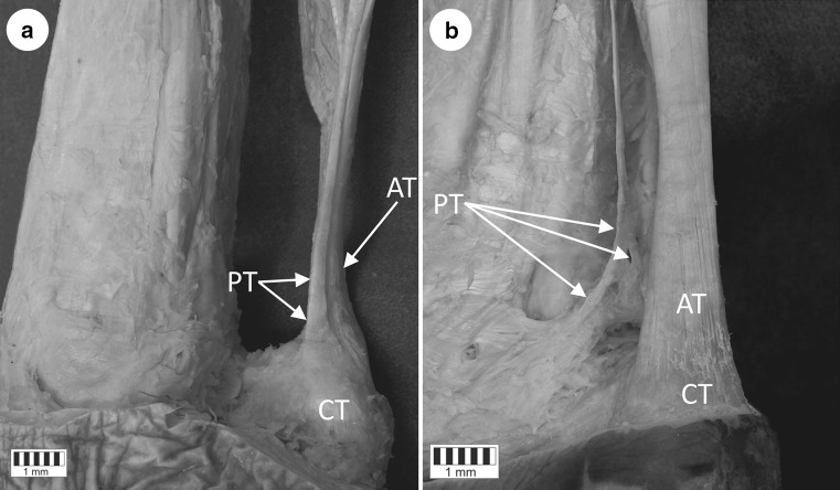
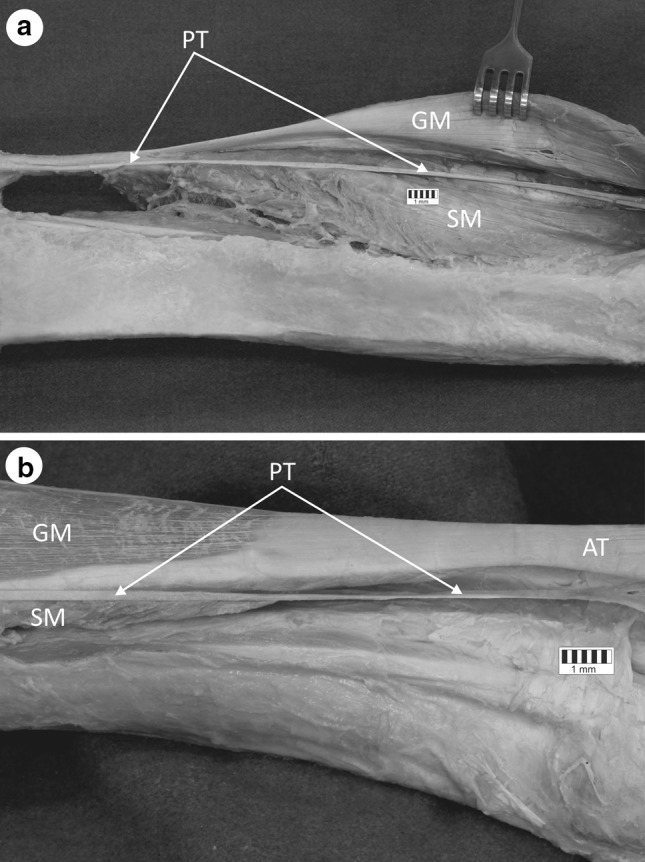
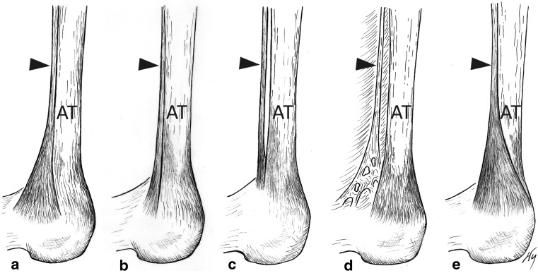
Five insertion types of the plantaris tendon were identified in relation to the calcaneal tendon: four with their insertion on the calcaneal tuberosity (Types 1, 2, 3, 5), while the fifth (Type 4) had its insertion in the crural fascia. In addition, two variants of the course of the plantaris tendon were identified, the most common being termed Variant A, in which the tendon crosses the space between the gastrocnemius and the soleus muscles, thus reaching the medial crural region, and is located on the medial side of the calcaneal tendon (84 % cases). The course of the Variant B is similar to the course of the Variant A, but upon leaving the space located between the gastrocnemius and soleus muscle, it turned to the medial crural region and ran directly anterior to the calcaneal tendon (12 %). The plantaris muscle was found to be absent in two lower limbs (4 %). The most frequent insertion type of the plantaris tendon into the calcaneal tuberosity is fan-shaped, occurring on the medial side of the Achilles tendon (Type 1-44 % cases).
The course of the plantaris tendon and its mobility range in relation to the calcaneal tendon may be likely to affect the occurrence of pains in the lower medial part of the leg (Achilles tendinopathy).
跟腱病是近年来临床上观察到的一个重要的下肢问题。疼痛背后的位置和机制均尚未得到充分解释。患者经常在跟腱内侧、跟骨结节上方2至7厘米处感到疼痛,这可能表明跖肌腱起着重要作用。本研究的目的是确定跖肌腱走行与跟腱之间的解剖关系,以及跖肌腱的附着类型。
对固定于10%甲醛溶液中的50条随机选取的下肢(23条左侧和27条右侧)进行检测。
确定了跖肌腱相对于跟腱的五种附着类型:四种附着于跟骨结节(1型、2型、3型、5型),而第五种(4型)附着于小腿筋膜。此外,还确定了跖肌腱走行的两种变异类型,最常见的称为变异A,其中该肌腱穿过腓肠肌和比目鱼肌之间的间隙,从而到达小腿内侧区域,并位于跟腱内侧(84%的病例)。变异B的走行与变异A相似,但在离开腓肠肌和比目鱼肌之间的间隙后,转向小腿内侧区域,并直接走行于跟腱前方(12%)。在两条下肢(4%)中发现无跖肌。跖肌腱最常见的附着于跟骨结节的类型呈扇形,发生于跟腱内侧(1型 - 44%的病例)。
跖肌腱的走行及其相对于跟腱的活动范围可能会影响小腿内侧下部疼痛(跟腱病)的发生。