• 文献检索
  • 文档翻译
  • 深度研究
  • 学术资讯
  • Suppr Zotero 插件Zotero 插件
  • 邀请有礼
  • 套餐&价格
  • 历史记录
应用&插件
Suppr Zotero 插件Zotero 插件浏览器插件Mac 客户端Windows 客户端微信小程序
定价
高级版会员购买积分包购买API积分包
服务
文献检索文档翻译深度研究API 文档MCP 服务
关于我们
关于 Suppr公司介绍联系我们用户协议隐私条款
关注我们

Suppr 超能文献

核心技术专利:CN118964589B侵权必究
粤ICP备2023148730 号-1Suppr @ 2026

文献检索

告别复杂PubMed语法,用中文像聊天一样搜索,搜遍4000万医学文献。AI智能推荐,让科研检索更轻松。

立即免费搜索

文件翻译

保留排版,准确专业,支持PDF/Word/PPT等文件格式,支持 12+语言互译。

免费翻译文档

深度研究

AI帮你快速写综述,25分钟生成高质量综述,智能提取关键信息,辅助科研写作。

立即免费体验

基于磁共振图像的合成 CT 用于调强适形前列腺治疗计划和锥形束 CT 图像引导定位。

MR image-based synthetic CT for IMRT prostate treatment planning and CBCT image-guided localization.

机构信息

Wuhan University; Beaumont Health System.

出版信息

J Appl Clin Med Phys. 2016 May 8;17(3):236-245. doi: 10.1120/jacmp.v17i3.6065.

DOI:10.1120/jacmp.v17i3.6065
PMID:27167281
原文链接:https://pmc.ncbi.nlm.nih.gov/articles/PMC5690904/
Abstract

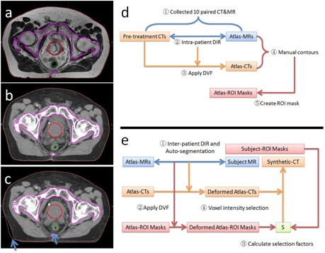
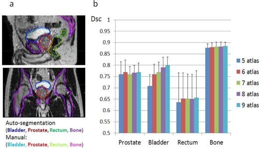
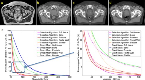
The purpose of this study was to propose and evaluate a method of creating a synthetic CT (S-CT) from MRI simulation for dose calculation and daily CBCT localization. A pair of MR and CT images was obtained in the same day from each of 10 prostate patients. The pair of MR and CT images was preregistered using the deformable image registration (DIR). Using the corresponding displacement vector field (atlas-DVF), the CT image was deformed to the MR image to create an atlas MR-CT pair. Regions of interest (ROI) on the atlas MR-CT pair were delineated and used to create atlas-ROI masks. 'Leave-one-out' test (one pair of MR and CT was used as subject-MR and subject-CT for evaluation, and the remaining 9 pairs were in the atlas library) was performed. For a subject-MR, autosegmentation and DVFs were generated using DIR between the subject-MR and the 9 atlas-MRs. An S-CT was then generated using the corresponding 9 paired atlas-CTs, the 9 atlas-DVFs and the corresponding atlas-ROI masks. The total 10 S-CTs were evaluated using the Hounsfield unit (HU), the calculated dose distribution, and the auto bony registration to daily CBCT images with respect to the 10 subject-CTs. HU differences (mean ± STD) were (2.4 ± 25.23), (1.18 ± 39.49), (32.46 ± 81.9), (0.23 ± 40.13), and (3.74 ± 144.76) for prostate, bladder, rectal wall, soft tissue outside all ROIs, and bone, respectively. The discrepancy of dose-volume param-eters calculated using the S-CT for treatment planning was small (≤ 0.22% with 95% confidence). Gamma pass rate (2% & 2 mm) was higher than 99.86% inside PTV and 98.45% inside normal structures. Using the 10 S-CTs as the reference CT for daily CBCT localization achieved the similar results compared to using the subject-CT. The translational vector differences were within 1.08 mm (0.37 ± 0.23 mm), and the rotational differences were within 1.1° in all three directions. S-CT created from a simulation MR image using the proposed approach with the preconstructed atlas library can replace the planning CT for dose calculation and daily CBCT image guidance.

摘要

本研究旨在提出并评估一种从 MRI 模拟中创建合成 CT(S-CT)以进行剂量计算和日常 CBCT 定位的方法。对 10 例前列腺患者的同一天内的一对 MR 和 CT 图像进行了采集。使用可变形图像配准(DIR)对 MR 和 CT 图像进行预配准。使用相应的位移向量场(图谱-DVF)将 CT 图像变形到 MR 图像,以创建图谱 MR-CT 对。在图谱 MR-CT 对上勾勒出感兴趣区域(ROI),并用于创建图谱 ROI 蒙版。进行了“留一法”测试(一对 MR 和 CT 被用作受试 MR 和受试 CT 进行评估,其余 9 对用于图谱库)。对于受试 MR,使用 DIR 在受试 MR 与 9 个图谱 MR 之间生成自动分割和 DVF。然后,使用相应的 9 对图谱 CT、9 个图谱 DVF 和相应的图谱 ROI 蒙版生成 S-CT。对 10 个受试 CT 的 10 个 S-CT 进行了基于体素 HU、计算剂量分布以及与日常 CBCT 图像的自动骨性配准的评估。HU 差异(均值±标准差)分别为前列腺(2.4±25.23)、膀胱(1.18±39.49)、直肠壁(32.46±81.9)、所有 ROI 外软组织(0.23±40.13)和骨骼(3.74±144.76)。使用 S-CT 进行治疗计划计算的剂量-体积参数差异较小(置信度为 95%时≤0.22%)。在 PTV 内的伽玛通过率(2%和 2mm)高于 99.86%,在正常结构内的通过率高于 98.45%。使用 10 个 S-CT 作为日常 CBCT 定位的参考 CT 可获得与使用受试 CT 相似的结果。平移向量差异在 1.08mm 以内(0.37±0.23mm),三个方向的旋转差异均在 1.1°以内。使用预先构建的图谱库从模拟 MR 图像创建的 S-CT 可以替代计划 CT 进行剂量计算和日常 CBCT 图像引导。

https://cdn.ncbi.nlm.nih.gov/pmc/blobs/f4d0/5690904/dac285c324d7/ACM2-17-236-g004.jpg
https://cdn.ncbi.nlm.nih.gov/pmc/blobs/f4d0/5690904/d86121d33842/ACM2-17-236-g001.jpg
https://cdn.ncbi.nlm.nih.gov/pmc/blobs/f4d0/5690904/d7b00aad4558/ACM2-17-236-g002.jpg
https://cdn.ncbi.nlm.nih.gov/pmc/blobs/f4d0/5690904/9dc160418be2/ACM2-17-236-g003.jpg
https://cdn.ncbi.nlm.nih.gov/pmc/blobs/f4d0/5690904/dac285c324d7/ACM2-17-236-g004.jpg

相似文献

1
MR image-based synthetic CT for IMRT prostate treatment planning and CBCT image-guided localization.基于磁共振图像的合成 CT 用于调强适形前列腺治疗计划和锥形束 CT 图像引导定位。
J Appl Clin Med Phys. 2016 May 8;17(3):236-245. doi: 10.1120/jacmp.v17i3.6065.
2
Validation of a deformable image registration technique for cone beam CT-based dose verification.基于锥形束CT的剂量验证中可变形图像配准技术的验证
Med Phys. 2015 Jan;42(1):196-205. doi: 10.1118/1.4903292.
3
Dosimetric and volumetric changes in the rectum and bladder in patients receiving CBCT-guided prostate IMRT: analysis based on daily CBCT dose calculation.接受CBCT引导的前列腺调强放疗患者直肠和膀胱的剂量学及容积变化:基于每日CBCT剂量计算的分析
J Appl Clin Med Phys. 2016 Nov 8;17(6):107-117. doi: 10.1120/jacmp.v17i6.6207.
4
Evaluation of online/offline image guidance/adaptation approaches for prostate cancer radiation therapy.前列腺癌放射治疗的在线/离线图像引导/适配方法评估
Int J Radiat Oncol Biol Phys. 2015 Apr 1;91(5):1026-33. doi: 10.1016/j.ijrobp.2014.12.043.
5
An atlas-based electron density mapping method for magnetic resonance imaging (MRI)-alone treatment planning and adaptive MRI-based prostate radiation therapy.基于图谱的磁共振成像(MRI)单模态治疗计划电子密度映射方法及其在自适应 MRI 引导前列腺放射治疗中的应用。
Int J Radiat Oncol Biol Phys. 2012 May 1;83(1):e5-11. doi: 10.1016/j.ijrobp.2011.11.056. Epub 2012 Feb 11.
6
Investigating the generalisation of an atlas-based synthetic-CT algorithm to another centre and MR scanner for prostate MR-only radiotherapy.研究基于图谱的合成 CT 算法在另一个中心和磁共振扫描仪上对前列腺磁共振-only 放疗的泛化能力。
Phys Med Biol. 2017 Nov 21;62(24):N548-N560. doi: 10.1088/1361-6560/aa9676.
7
Automatic Substitute Computed Tomography Generation and Contouring for Magnetic Resonance Imaging (MRI)-Alone External Beam Radiation Therapy From Standard MRI Sequences.基于标准 MRI 序列的 MRI 引导外束放射治疗中自动替代 CT 生成和勾画。
Int J Radiat Oncol Biol Phys. 2015 Dec 1;93(5):1144-53. doi: 10.1016/j.ijrobp.2015.08.045. Epub 2015 Sep 5.
8
Determination of optimal PTV margin for patients receiving CBCT-guided prostate IMRT: comparative analysis based on CBCT dose calculation with four different margins.接受CBCT引导的前列腺调强放疗患者最佳计划靶区(PTV)边界的确定:基于四种不同边界的CBCT剂量计算的对比分析
J Appl Clin Med Phys. 2015 Nov 8;16(6):252–262. doi: 10.1120/jacmp.v16i6.5691.
9
Dosimetric evaluation of an atlas-based synthetic CT generation approach for MR-only radiotherapy of pelvis anatomy.基于图谱的合成CT生成方法在骨盆解剖结构仅磁共振放疗中的剂量学评估
J Appl Clin Med Phys. 2019 Jan;20(1):101-109. doi: 10.1002/acm2.12501. Epub 2018 Nov 26.
10
An adaptive MR-CT registration method for MRI-guided prostate cancer radiotherapy.一种用于MRI引导的前列腺癌放疗的自适应MR-CT配准方法。
Phys Med Biol. 2015 Apr 7;60(7):2837-51. doi: 10.1088/0031-9155/60/7/2837. Epub 2015 Mar 17.

引用本文的文献

1
Generation of tissues outside the field of view (FOV) of radiation therapy simulation imaging based on machine learning and patient body outline (PBO).基于机器学习和患者身体轮廓(PBO)生成放射治疗模拟成像视野(FOV)之外的组织。
Radiat Oncol. 2024 Jan 25;19(1):15. doi: 10.1186/s13014-023-02384-4.
2
Prostate volume analysis in image registration for prostate cancer care: a verification study.图像配准中前列腺体积分析在前列腺癌治疗中的应用:验证性研究。
Phys Eng Sci Med. 2023 Dec;46(4):1791-1802. doi: 10.1007/s13246-023-01342-4. Epub 2023 Oct 11.
3
Synthetic computed tomography generation for abdominal adaptive radiotherapy using low-field magnetic resonance imaging.

本文引用的文献

1
Quantitative characterizations of ultrashort echo (UTE) images for supporting air-bone separation in the head.用于支持头部气骨分离的超短回波(UTE)图像的定量表征。
Phys Med Biol. 2015 Apr 7;60(7):2869-80. doi: 10.1088/0031-9155/60/7/2869. Epub 2015 Mar 17.
2
Generating patient specific pseudo-CT of the head from MR using atlas-based regression.使用基于图谱的回归方法从磁共振成像(MR)生成头部患者特异性的伪计算机断层扫描(CT)。
Phys Med Biol. 2015 Jan 21;60(2):825-39. doi: 10.1088/0031-9155/60/2/825. Epub 2015 Jan 7.
3
A unifying probabilistic Bayesian approach to derive electron density from MRI for radiation therapy treatment planning.
利用低场磁共振成像生成用于腹部自适应放疗的合成计算机断层扫描
Phys Imaging Radiat Oncol. 2023 Feb 23;25:100425. doi: 10.1016/j.phro.2023.100425. eCollection 2023 Jan.
4
A convolutional neural network model for EPID-based non-transit dosimetry.基于 EPID 的非透射剂量学的卷积神经网络模型。
J Appl Clin Med Phys. 2023 Jun;24(6):e13923. doi: 10.1002/acm2.13923. Epub 2023 Mar 2.
5
Dosimetric Impact of Intrafraction Prostate Motion and Interfraction Anatomical Changes in Dose-Escalated Linac-Based SBRT.基于直线加速器的剂量递增立体定向体部放疗中分次内前列腺运动和分次间解剖结构变化的剂量学影响
Cancers (Basel). 2023 Feb 10;15(4):1153. doi: 10.3390/cancers15041153.
6
Synthetic digital reconstructed radiographs for MR-only robotic stereotactic radiation therapy: A proof of concept.仅磁共振机器人立体定向放疗的合成数字重建射线照片:概念验证。
Comput Biol Med. 2021 Nov;138:104917. doi: 10.1016/j.compbiomed.2021.104917. Epub 2021 Oct 4.
7
The accuracy of Magnetic Resonance - Cone Beam Computed Tomography soft-tissue matching for prostate radiotherapy.磁共振-锥形束计算机断层扫描软组织匹配在前列腺癌放疗中的准确性
Phys Imaging Radiat Oncol. 2019 Dec 5;12:49-55. doi: 10.1016/j.phro.2019.11.005. eCollection 2019 Oct.
8
Dosimetric evaluation of synthetic CT generated with GANs for MRI-only proton therapy treatment planning of brain tumors.基于生成对抗网络(GANs)生成的合成CT在脑肿瘤仅磁共振成像质子治疗计划中的剂量学评估
J Appl Clin Med Phys. 2020 May;21(5):76-86. doi: 10.1002/acm2.12856. Epub 2020 Mar 26.
9
Introduction of a Simple Algorithm to Create Synthetic-computed Tomography of the Head from Magnetic Resonance Imaging.一种从磁共振成像创建头部合成计算机断层扫描的简单算法介绍。
J Med Signals Sens. 2019 Apr-Jun;9(2):123-129. doi: 10.4103/jmss.JMSS_26_18.
10
FMEA of MR-Only Treatment Planning in the Pelvis.骨盆仅磁共振治疗计划的失效模式与效应分析
Adv Radiat Oncol. 2018 Sep 7;4(1):168-176. doi: 10.1016/j.adro.2018.08.024. eCollection 2019 Jan-Mar.
一种用于从磁共振成像(MRI)中推导电子密度以进行放射治疗治疗计划的统一概率贝叶斯方法。
Phys Med Biol. 2014 Nov 7;59(21):6595-606. doi: 10.1088/0031-9155/59/21/6595. Epub 2014 Oct 16.
4
Feasibility of MRI-based reference images for image-guided radiotherapy of the pelvis with either cone-beam computed tomography or planar localization images.基于磁共振成像(MRI)的参考图像用于盆腔图像引导放射治疗(采用锥形束计算机断层扫描或平面定位图像)的可行性。
Acta Oncol. 2015 Jun;54(6):889-95. doi: 10.3109/0284186X.2014.958197. Epub 2014 Sep 18.
5
Counterpoint: Opportunities and challenges of a magnetic resonance imaging-only radiotherapy work flow.反驳观点:仅使用磁共振成像的放射治疗工作流程的机遇与挑战
Semin Radiat Oncol. 2014 Jul;24(3):175-80. doi: 10.1016/j.semradonc.2014.02.005.
6
MRI-based treatment planning with pseudo CT generated through atlas registration.基于图谱配准生成的伪CT进行MRI引导的治疗计划
Med Phys. 2014 May;41(5):051711. doi: 10.1118/1.4873315.
7
A dual model HU conversion from MRI intensity values within and outside of bone segment for MRI-based radiotherapy treatment planning of prostate cancer.一种用于基于MRI的前列腺癌放射治疗计划的双模型HU转换,该转换基于骨段内外的MRI强度值。
Med Phys. 2014 Jan;41(1):011704. doi: 10.1118/1.4842575.
8
T1/T2*-weighted MRI provides clinically relevant pseudo-CT density data for the pelvic bones in MRI-only based radiotherapy treatment planning.T1/T2*-加权 MRI 可为基于 MRI 的放射治疗计划提供临床相关的骨盆伪 CT 密度数据。
Acta Oncol. 2013 Apr;52(3):612-8. doi: 10.3109/0284186X.2012.692883. Epub 2012 Jun 19.
9
Regression-Based Label Fusion for Multi-Atlas Segmentation.基于回归的标签融合用于多图谱分割
Conf Comput Vis Pattern Recognit Workshops. 2011 Jun 20:1113-1120. doi: 10.1109/CVPR.2011.5995382.
10
An atlas-based electron density mapping method for magnetic resonance imaging (MRI)-alone treatment planning and adaptive MRI-based prostate radiation therapy.基于图谱的磁共振成像(MRI)单模态治疗计划电子密度映射方法及其在自适应 MRI 引导前列腺放射治疗中的应用。
Int J Radiat Oncol Biol Phys. 2012 May 1;83(1):e5-11. doi: 10.1016/j.ijrobp.2011.11.056. Epub 2012 Feb 11.