Wei Hongjiang, Xie Luke, Dibb Russell, Li Wei, Decker Kyle, Zhang Yuyao, Johnson G Allan, Liu Chunlei
Brain Imaging and Analysis Center, Duke University, Durham, NC 27705, USA.
Utah Center for Advanced Imaging Research, Department of Radiology, University of Utah, Salt Lake City, UT 84108, USA.
Neuroimage. 2016 Aug 15;137:107-115. doi: 10.1016/j.neuroimage.2016.05.033. Epub 2016 May 12.
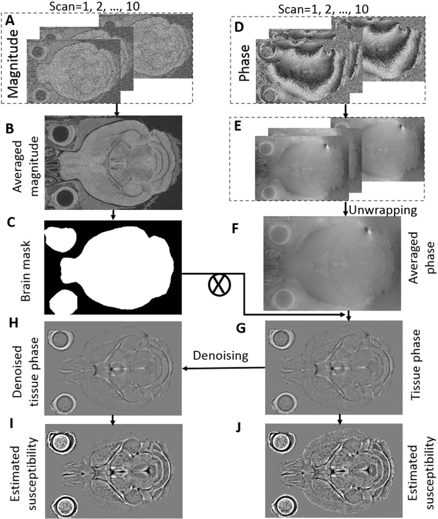
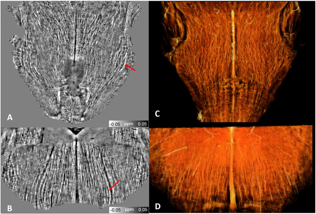
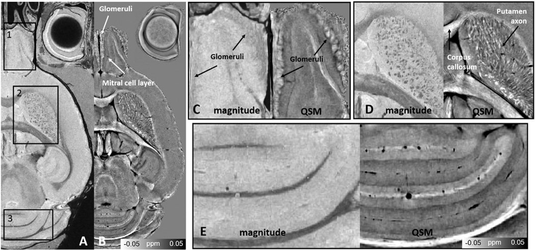
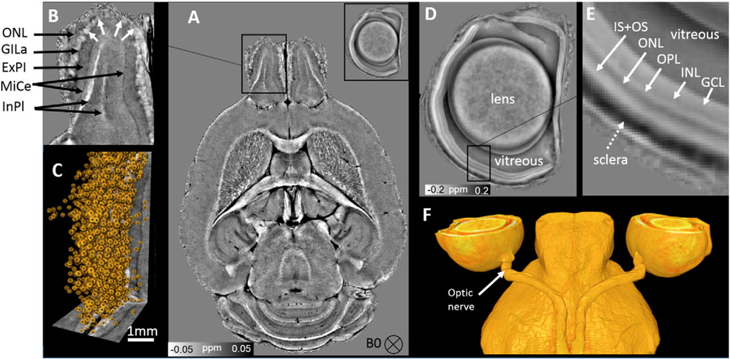
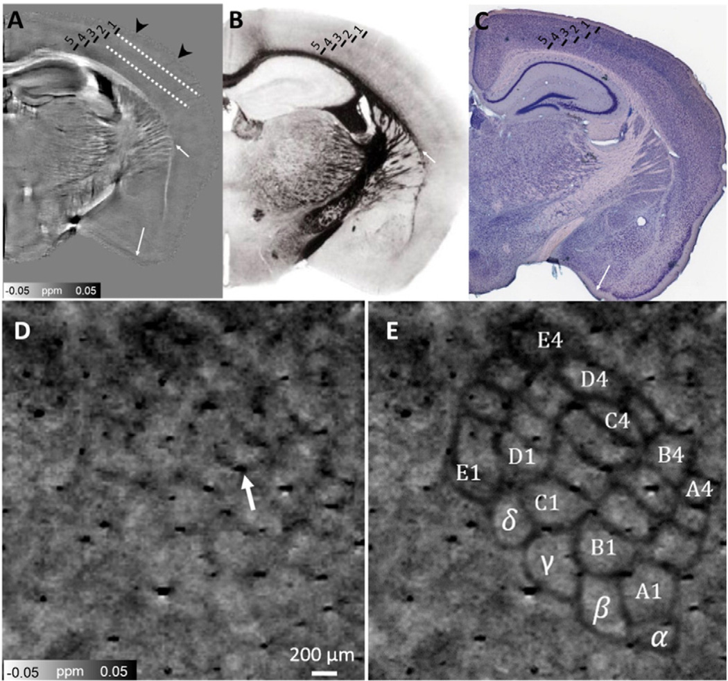
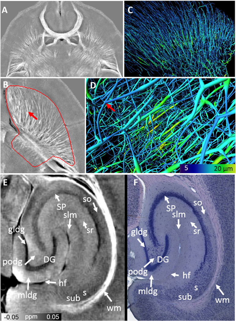
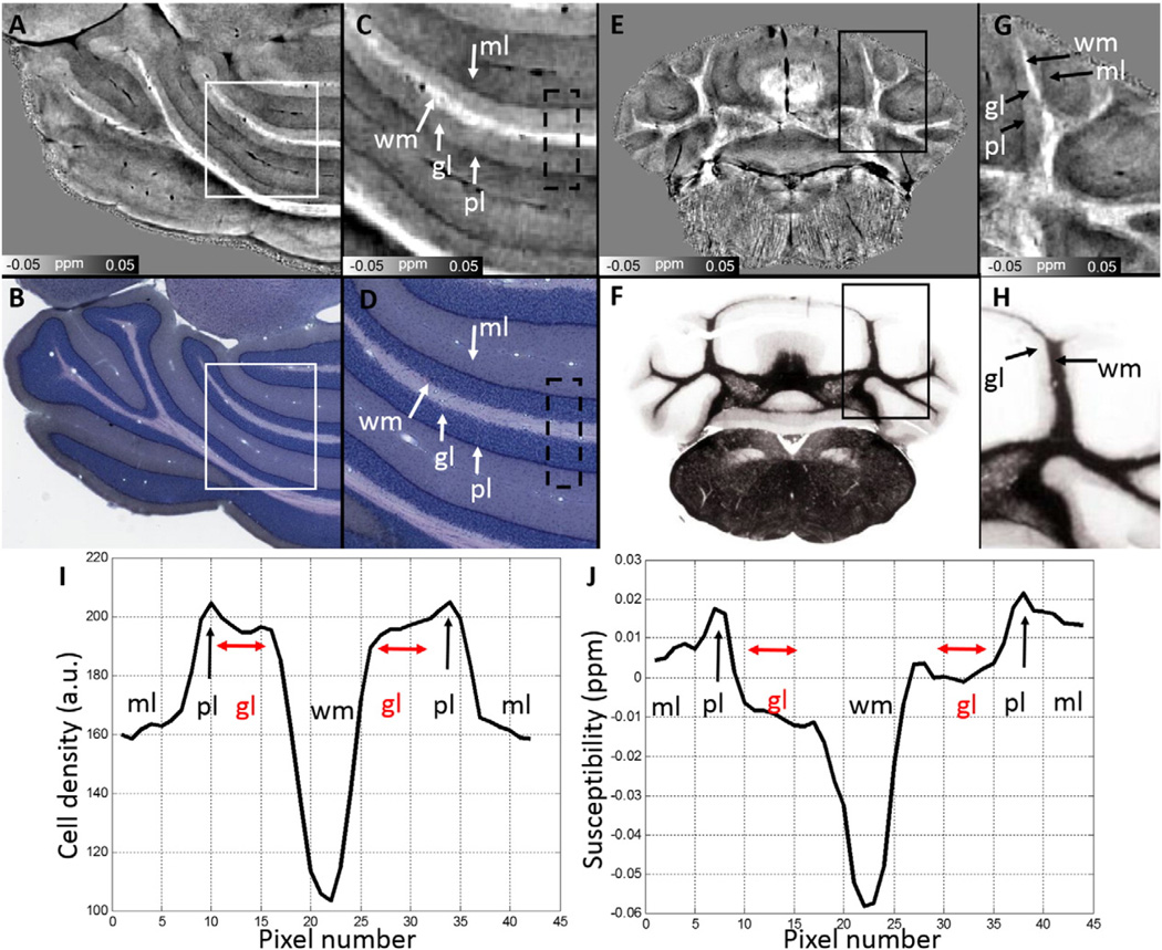
The proper microstructural arrangement of complex neural structures is essential for establishing the functional circuitry of the brain. We present an MRI method to resolve tissue microstructure and infer brain cytoarchitecture by mapping the magnetic susceptibility in the brain at high resolution. This is possible because of the heterogeneous magnetic susceptibility created by varying concentrations of lipids, proteins and irons from the cell membrane to cytoplasm. We demonstrate magnetic susceptibility maps at a nominal resolution of 10-μm isotropic, approaching the average cell size of a mouse brain. The maps reveal many detailed structures including the retina cell layers, olfactory sensory neurons, barrel cortex, cortical layers, axonal fibers in white and gray matter. Olfactory glomerulus density is calculated and structural connectivity is traced in the optic nerve, striatal neurons, and brainstem nerves. The method is robust and can be readily applied on MRI scanners at or above 7T.
复杂神经结构的适当微观结构排列对于建立大脑的功能回路至关重要。我们提出了一种磁共振成像(MRI)方法,通过高分辨率绘制大脑中的磁化率来解析组织微观结构并推断脑细结构。这是可行的,因为从细胞膜到细胞质,脂质、蛋白质和铁的浓度变化会产生不均匀的磁化率。我们展示了名义分辨率为各向同性10μm的磁化率图,接近小鼠大脑的平均细胞大小。这些图揭示了许多详细结构,包括视网膜细胞层、嗅觉感觉神经元、桶状皮质、皮质层、白质和灰质中的轴突纤维。计算了嗅觉小球密度,并追踪了视神经、纹状体神经元和脑干神经中的结构连接性。该方法稳健,可轻松应用于7T及以上的MRI扫描仪。