Gutierrez-Mecinas Maria, Kuehn Emily D, Abraira Victoria E, Polgár Erika, Watanabe Masahiko, Todd Andrew J
Institute of Neuroscience and Psychology, College of Medical, Veterinary and Life Sciences, University of Glasgow, Glasgow G12 8QQ, UK.
Department of Neurobiology, Howard Hughes Medical Institute, Harvard Medical School, Boston, MA 02115, USA.
Neuroscience. 2016 Aug 4;329:171-81. doi: 10.1016/j.neuroscience.2016.05.009. Epub 2016 May 13.
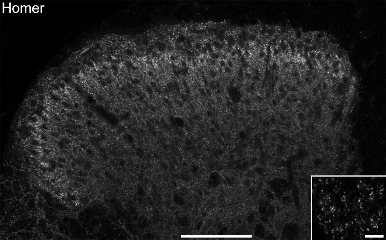
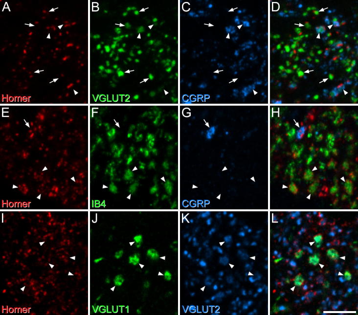
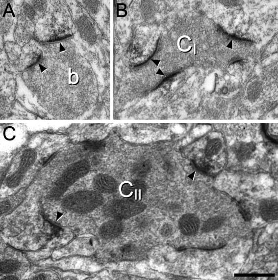
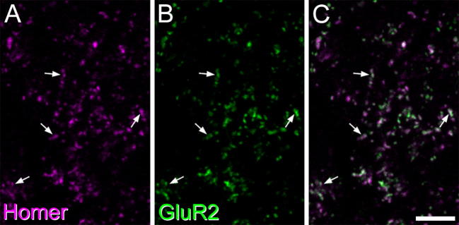
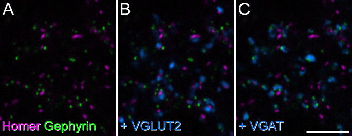
The spinal dorsal horn processes somatosensory information before conveying it to the brain. The neuronal organization of the dorsal horn is still poorly understood, although recent studies have defined several distinct populations among the interneurons, which account for most of its constituent neurons. All primary afferents, and the great majority of neurons in laminae I-III are glutamatergic, and a major factor limiting our understanding of the synaptic circuitry has been the difficulty in identifying glutamatergic synapses with light microscopy. Although there are numerous potential targets for antibodies, these are difficult to visualize with immunocytochemistry, because of protein cross-linking following tissue fixation. Although this can be overcome by antigen retrieval methods, these lead to difficulty in detecting other antigens. The aim of this study was to test whether the postsynaptic protein Homer can be used to reveal glutamatergic synapses in the dorsal horn. Immunostaining for Homer gave punctate labeling when viewed by confocal microscopy, and this was restricted to synapses at the ultrastructural level. We found that Homer puncta were colocalized with the AMPA receptor GluR2 subunit, but not with the inhibitory synapse-associated protein gephyrin. We also examined several populations of glutamatergic axons and found that most boutons were in contact with at least one Homer punctum. These results suggest that Homer antibodies can be used to reveal the great majority of glutamatergic synapses without antigen retrieval. This will be of considerable value in tracing synaptic circuits, and also in investigating plasticity of glutamatergic synapses in pain states.
脊髓背角在将躯体感觉信息传递至大脑之前会对其进行处理。尽管最近的研究已经明确了中间神经元中的几个不同群体,而中间神经元构成了背角的大部分神经元,但背角的神经元组织仍未被充分了解。所有的初级传入神经以及板层I-III中的绝大多数神经元都是谷氨酸能的,而限制我们对突触回路理解的一个主要因素是难以通过光学显微镜识别谷氨酸能突触。尽管有许多抗体的潜在靶点,但由于组织固定后蛋白质交联,这些靶点很难通过免疫细胞化学进行可视化。尽管这可以通过抗原修复方法克服,但这些方法会导致难以检测其他抗原。本研究的目的是测试突触后蛋白荷马(Homer)是否可用于揭示背角中的谷氨酸能突触。通过共聚焦显微镜观察时,对荷马的免疫染色呈现点状标记,且在超微结构水平上这种标记仅限于突触。我们发现荷马斑点与AMPA受体GluR2亚基共定位,但与抑制性突触相关蛋白gephyrin不共定位。我们还检查了几个谷氨酸能轴突群体,发现大多数轴突终扣至少与一个荷马斑点接触。这些结果表明,无需抗原修复,荷马抗体可用于揭示绝大多数谷氨酸能突触。这对于追踪突触回路以及研究疼痛状态下谷氨酸能突触的可塑性将具有相当大的价值。