Lai Priscilla, Tarapacki Christine, Tran William T, El Kaffas Ahmed, Lee Justin, Hupple Clinton, Iradji Sarah, Giles Anoja, Al-Mahrouki Azza, Czarnota Gregory J
Imaging Research, Sunnybrook Health Sciences Centre, Toronto, Ontario, Canada; Department of Medical Biophysics, University of Toronto, Toronto, Ontario, Canada.
Radiation Oncology, Sunnybrook Health Sciences Centre, Toronto, Ontario, Canada; Department of Radiation Oncology, University of Toronto, Toronto, Ontario, Canada; Imaging Research, Sunnybrook Health Sciences Centre, Toronto, Ontario, Canada.
Oncoscience. 2016 Mar 24;3(3-4):98-108. doi: 10.18632/oncoscience.299. eCollection 2016.
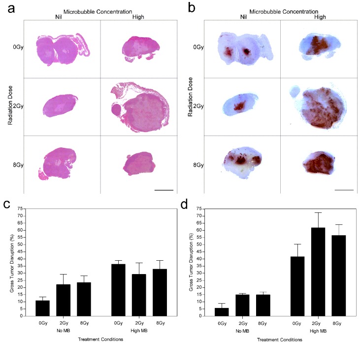
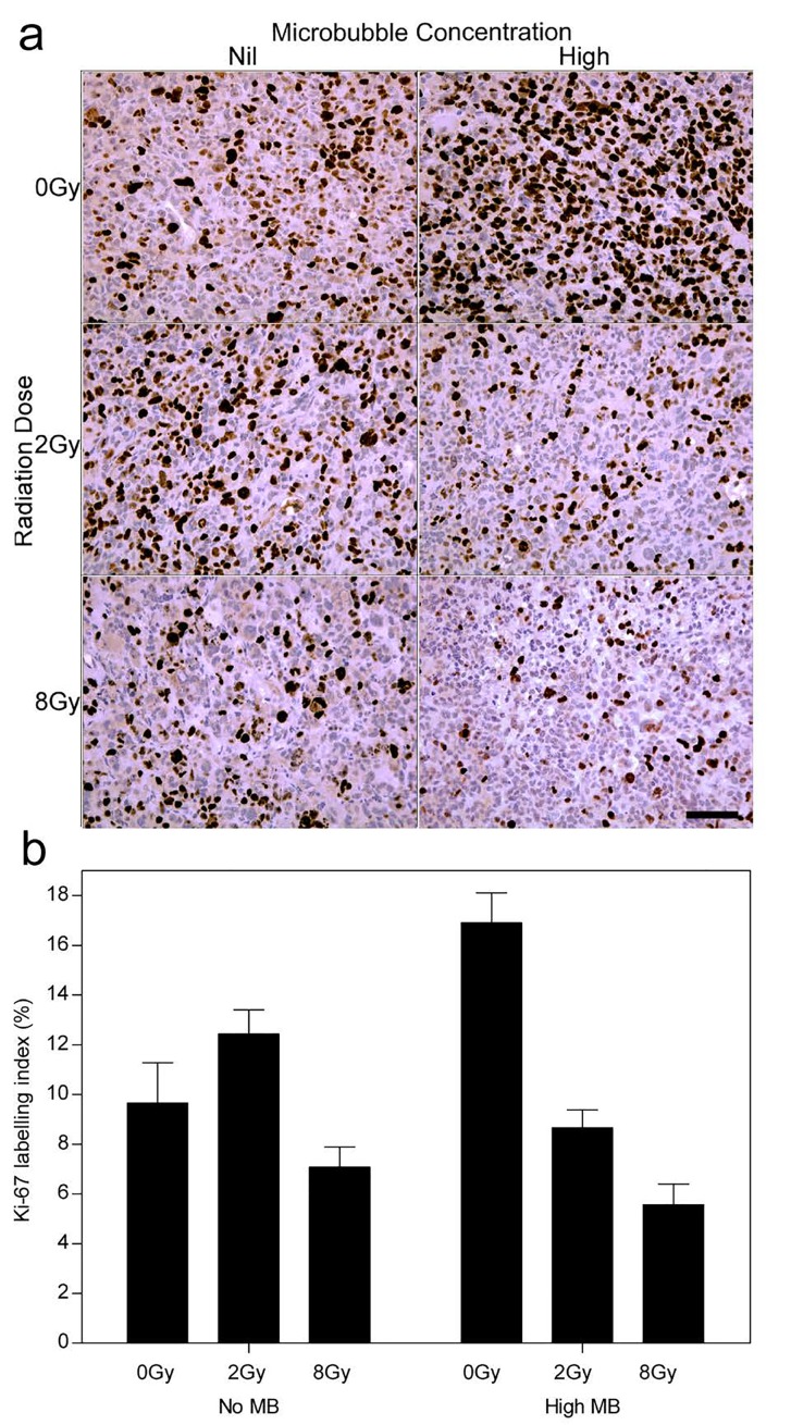
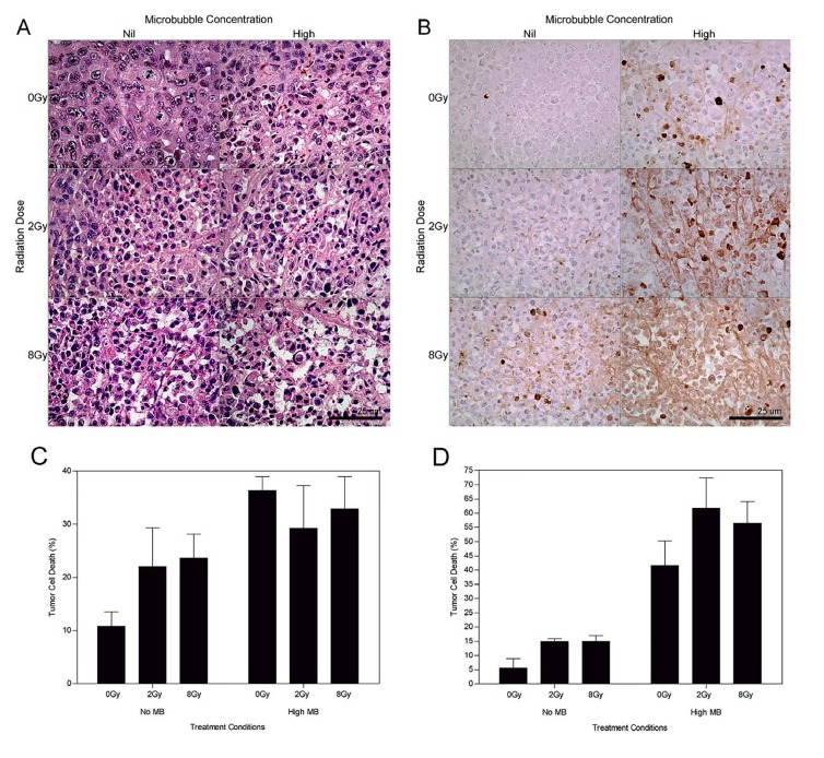
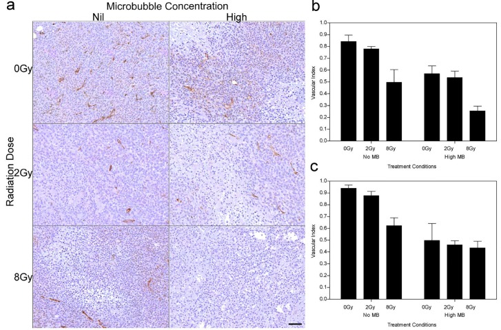
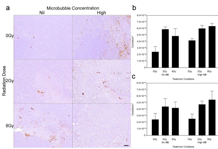
Acoustically stimulated microbubbles have been demonstrated to perturb endothelial cells of the vasculature resulting in biological effects. In the present study, vascular and tumor response to ultrasound-stimulated microbubble and radiation treatment was investigated in vivo to identify effects on the blood vessel endothelium. Mice bearing breast cancer tumors (MDA-MB-231) were exposed to ultrasound after intravenous injection of microbubbles at different concentrations, and radiation at different doses (0, 2, and 8 Gy). Mice were sacrificed 12 and 24 hours after treatment for histopathological analysis. Tumor growth delay was assessed for up to 28 days after treatment. The results demonstrated additive antitumor and antivascular effects when ultrasound stimulated microbubbles were combined with radiation. Results indicated tumor cell apoptosis, vascular leakage, a decrease in tumor vasculature, a delay in tumor growth and an overall tumor disruption. When coupled with radiation, ultrasound-stimulated microbubbles elicited synergistic anti-tumor and antivascular effects by acting as a radioenhancing agent in breast tumor blood vessels. The present study demonstrates ultrasound driven microbubbles as a novel form of targeted antiangiogenic therapy in a breast cancer xenograft model that can potentiate additive effects to radiation in vivo.
声学刺激的微泡已被证明会干扰脉管系统的内皮细胞,从而产生生物学效应。在本研究中,在体内研究了血管和肿瘤对超声刺激微泡及放射治疗的反应,以确定对血管内皮的影响。给携带乳腺癌肿瘤(MDA-MB-231)的小鼠静脉注射不同浓度的微泡后,再给予不同剂量(0、2和8 Gy)的辐射,然后对其进行超声照射。治疗后12小时和24小时处死小鼠进行组织病理学分析。在治疗后长达28天的时间里评估肿瘤生长延迟情况。结果表明,当超声刺激微泡与放射联合使用时,具有相加的抗肿瘤和抗血管作用。结果显示肿瘤细胞凋亡、血管渗漏、肿瘤脉管系统减少、肿瘤生长延迟以及整体肿瘤破坏。当与放射联合使用时,超声刺激微泡通过在乳腺肿瘤血管中充当放射增强剂,引发协同的抗肿瘤和抗血管作用。本研究证明,在乳腺癌异种移植模型中,超声驱动的微泡是一种新型的靶向抗血管生成疗法,可在体内增强对放射的相加作用。