Liang Po-Chin, Chen Yung-Chu, Chiang Chi-Feng, Mo Lein-Ray, Wei Shwu-Yuan, Hsieh Wen-Yuan, Lin Win-Li
Institute of Biomedical Engineering, College of Medicine, College of Engineering, National Taiwan University Hospital, Taipei, Taiwan; Department of Medical Imaging, National Taiwan University Hospital, Taipei, Taiwan.
Institute of Biomedical Engineering, College of Medicine, College of Engineering, National Taiwan University Hospital, Taipei, Taiwan; Biomedical Technology and Device Research Labs, Industrial Technology Research Institute, Hsinchu, Taiwan.
Int J Nanomedicine. 2016 May 12;11:2021-37. doi: 10.2147/IJN.S94139. eCollection 2016.
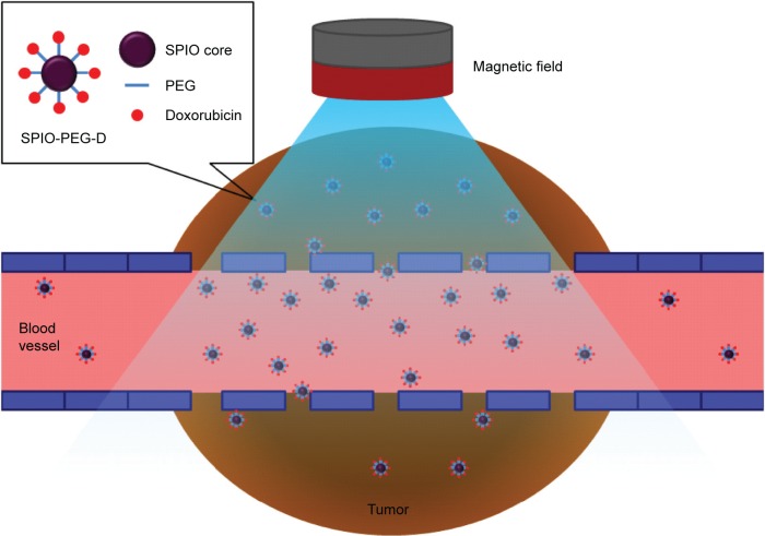
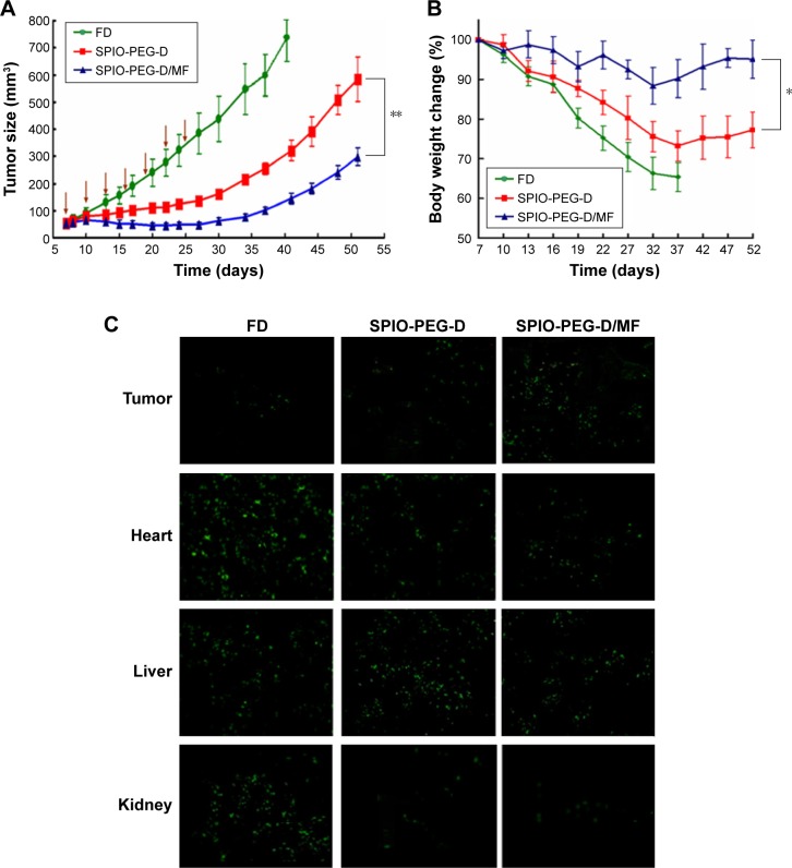
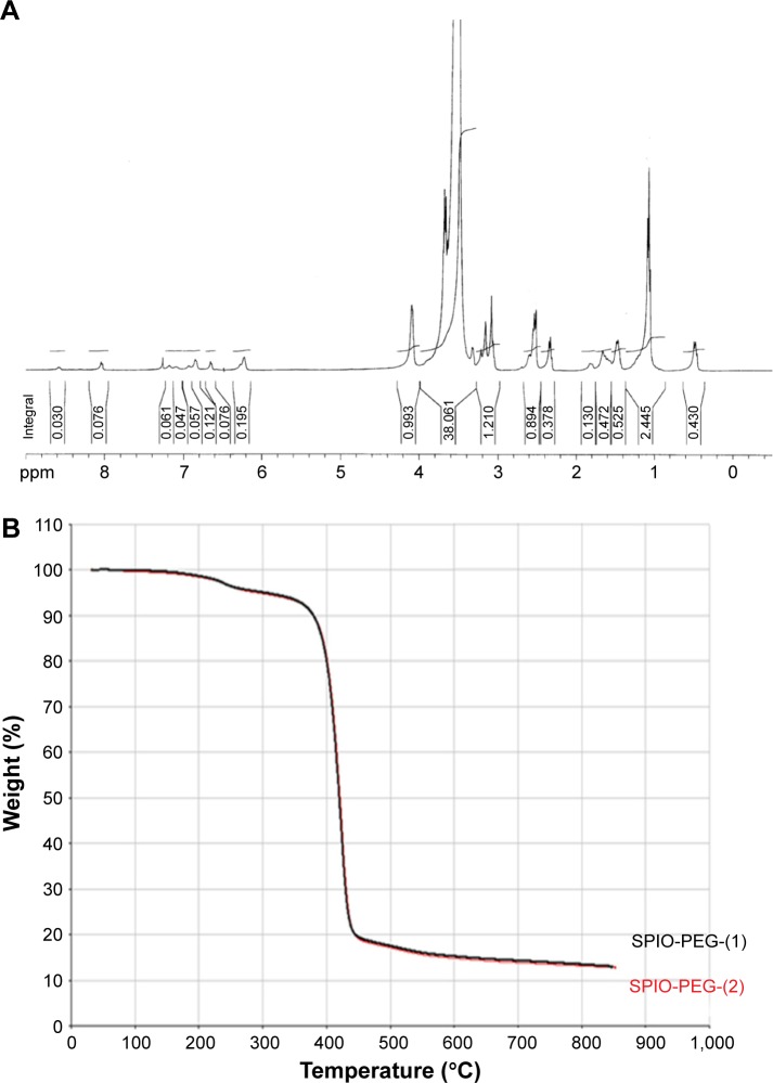
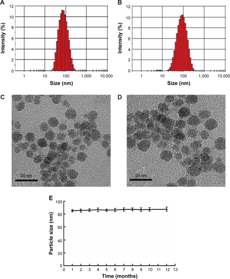
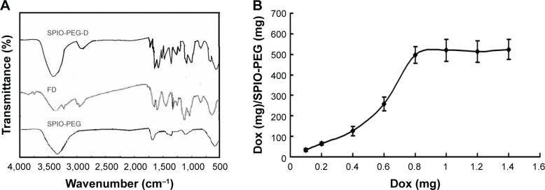
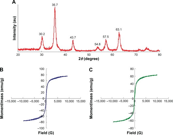
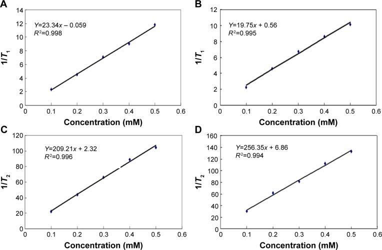
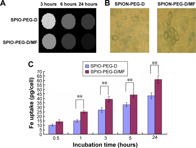
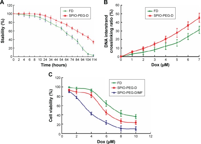
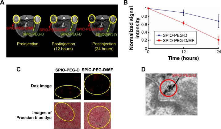
In this study, we developed functionalized superparamagnetic iron oxide (SPIO) nanoparticles consisting of a magnetic Fe3O4 core and a shell of aqueous stable polyethylene glycol (PEG) conjugated with doxorubicin (Dox) (SPIO-PEG-D) for tumor magnetic resonance imaging (MRI) enhancement and chemotherapy. The size of SPIO nanoparticles was ~10 nm, which was visualized by transmission electron microscope. The hysteresis curve, generated with vibrating-sample magnetometer, showed that SPIO-PEG-D was superparamagnetic with an insignificant hysteresis. The transverse relaxivity (r 2) for SPIO-PEG-D was significantly higher than the longitudinal relaxivity (r 1) (r 2/r 1 >10). The half-life of Dox in blood circulation was prolonged by conjugating Dox on the surface of SPIO with PEG to reduce its degradation. The in vitro experiment showed that SPIO-PEG-D could cause DNA crosslink more serious, resulting in a lower DNA expression and a higher cell apoptosis for HT-29 cancer cells. The Prussian blue staining study showed that the tumors treated with SPIO-PEG-D under a magnetic field had a much higher intratumoral iron density than the tumors treated with SPIO-PEG-D alone. The in vivo MRI study showed that the T2-weighted signal enhancement was stronger for the group under a magnetic field, indicating that it had a better accumulation of SPIO-PEG-D in tumor tissues. In the anticancer efficiency study for SPIO-PEG-D, the results showed that there was a significantly smaller tumor size for the group with a magnetic field than the group without. The in vivo experiments also showed that this drug delivery system combined with a local magnetic field could reduce the side effects of cardiotoxicity and hepatotoxicity. The results showed that the developed SPIO-PEG-D nanoparticles own a great potential for MRI-monitoring magnet-enhancing tumor chemotherapy.
在本研究中,我们开发了功能化的超顺磁性氧化铁(SPIO)纳米颗粒,其由磁性Fe3O4核以及与阿霉素(Dox)共轭的水性稳定聚乙二醇(PEG)壳层组成(SPIO-PEG-D),用于肿瘤磁共振成像(MRI)增强和化疗。SPIO纳米颗粒的尺寸约为10 nm,通过透射电子显微镜可见。用振动样品磁强计生成的磁滞曲线表明,SPIO-PEG-D具有超顺磁性且磁滞不明显。SPIO-PEG-D的横向弛豫率(r 2)显著高于纵向弛豫率(r 1)(r 2/r 1>10)。通过将Dox与PEG共轭在SPIO表面,延长了Dox在血液循环中的半衰期,以减少其降解。体外实验表明,SPIO-PEG-D可导致DNA交联更严重,导致HT-29癌细胞的DNA表达降低和细胞凋亡增加。普鲁士蓝染色研究表明,在磁场下用SPIO-PEG-D治疗的肿瘤比单独用SPIO-PEG-D治疗的肿瘤具有更高的瘤内铁密度。体内MRI研究表明,磁场下的组T2加权信号增强更强,表明其在肿瘤组织中对SPIO-PEG-D的蓄积更好。在SPIO-PEG-D的抗癌效率研究中,结果表明,有磁场组的肿瘤尺寸明显小于无磁场组。体内实验还表明,这种药物递送系统与局部磁场相结合可降低心脏毒性和肝毒性的副作用。结果表明,所开发的SPIO-PEG-D纳米颗粒在MRI监测磁增强肿瘤化疗方面具有巨大潜力。