Halin Bergström Sofia, Nilsson Maria, Adamo Hanibal, Thysell Elin, Jernberg Emma, Stattin Pär, Widmark Anders, Wikström Pernilla, Bergh Anders
Department of Medical Biosciences, Pathology, Umeå University, Umeå, Sweden.
Department of Surgical and Perioperative Sciences, Urology, Umeå University, Umeå, Sweden.
PLoS One. 2016 Jun 9;11(6):e0157280. doi: 10.1371/journal.pone.0157280. eCollection 2016.
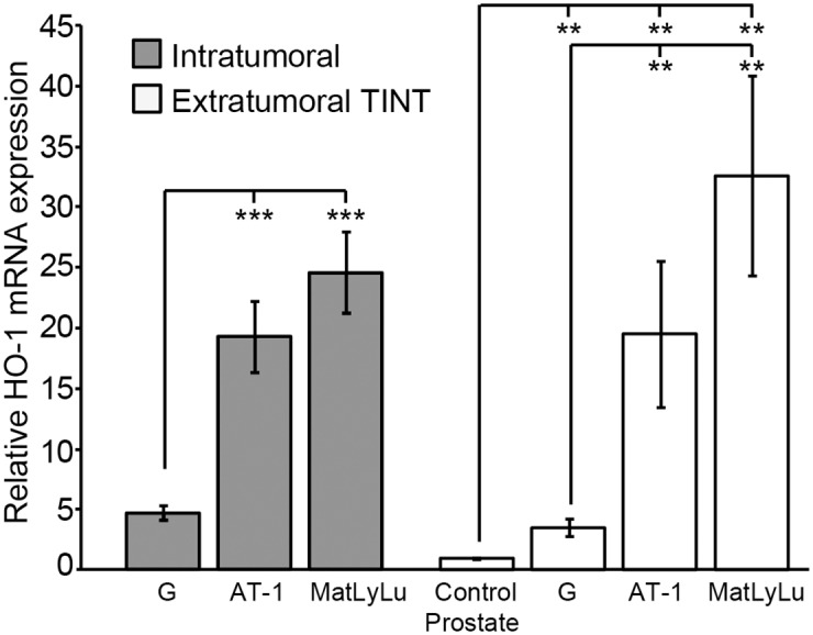
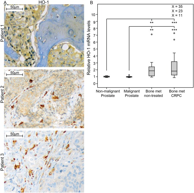
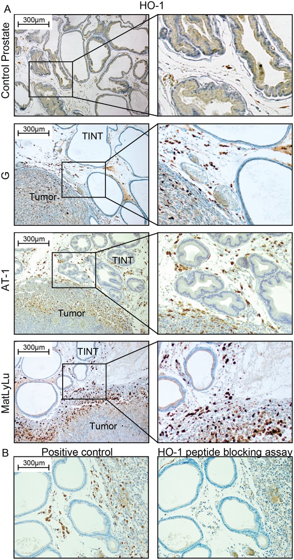
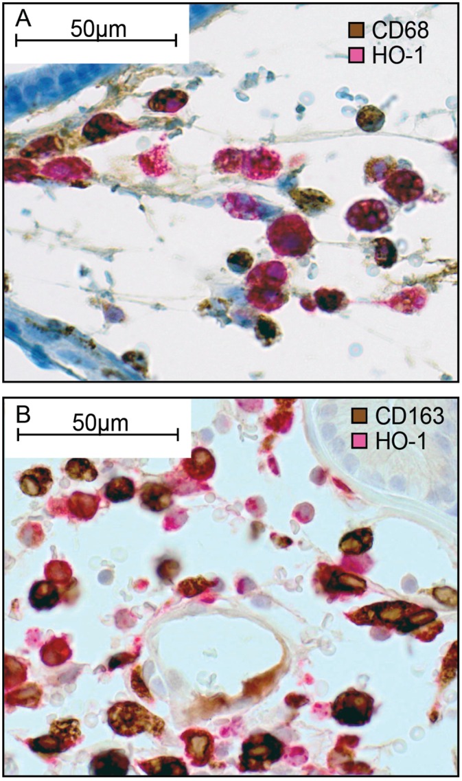
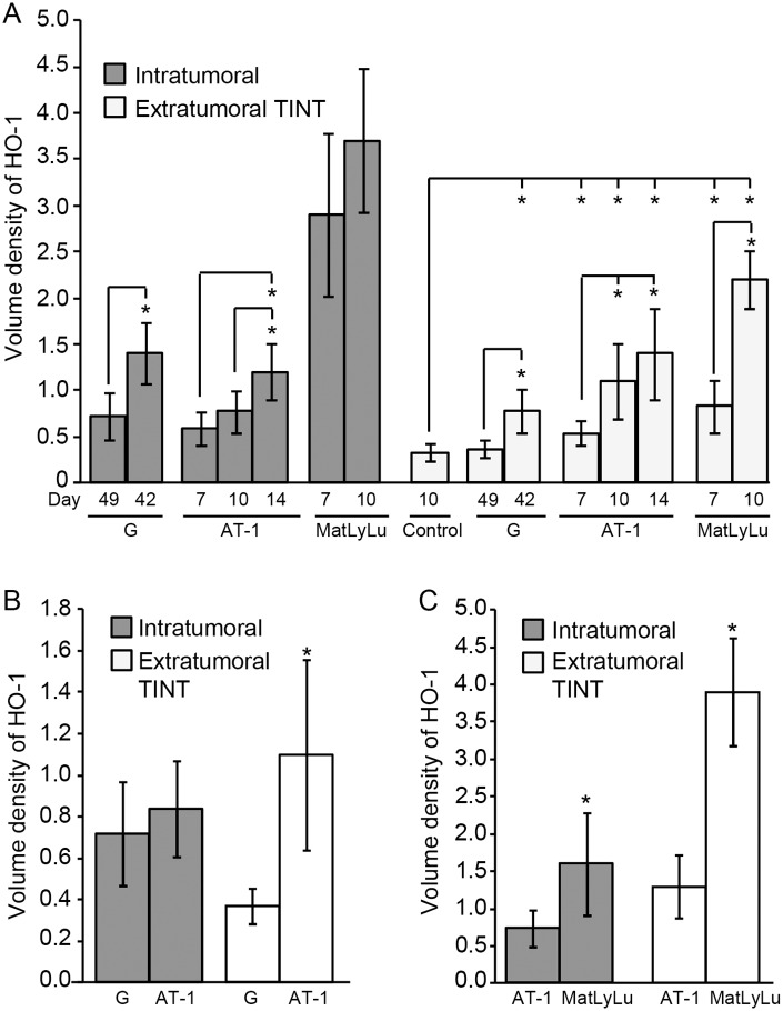
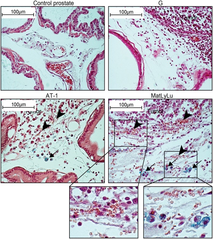
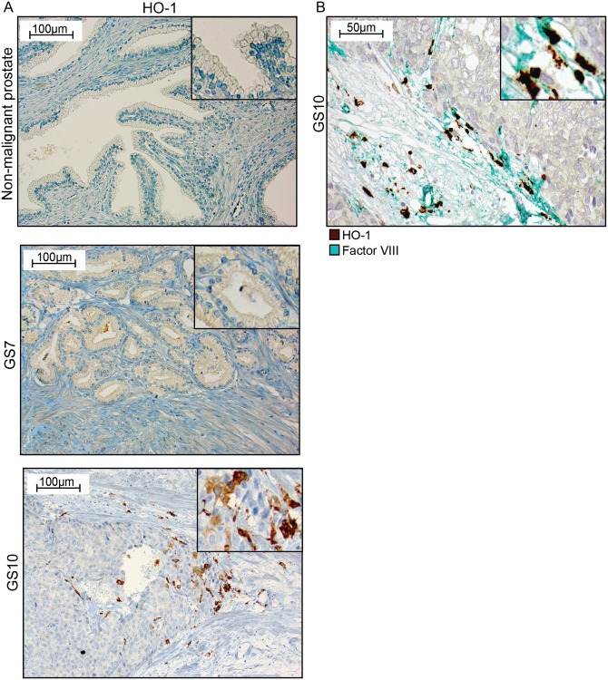
Aggressive tumors induce tumor-supporting changes in the benign parts of the prostate. One factor that has increased expression outside prostate tumors is hemoxygenase-1 (HO-1). To investigate HO-1 expression in more detail, we analyzed samples of tumor tissue and peritumoral normal prostate tissue from rats carrying cancers with different metastatic capacity, and human prostate cancer tissue samples from primary tumors and bone metastases. In rat prostate tumor samples, immunohistochemistry and quantitative RT-PCR showed that the main site of HO-1 synthesis was HO-1+ macrophages that accumulated in the tumor-bearing organ, and at the tumor-invasive front. Small metastatic tumors were considerably more effective in attracting HO-1+ macrophages than larger non-metastatic ones. In clinical samples, accumulation of HO-1+ macrophages was seen at the tumor invasive front, almost exclusively in high-grade tumors, and it correlated with the presence of bone metastases. HO-1+ macrophages, located at the tumor invasive front, were more abundant in bone metastases than in primary tumors. HO-1 expression in bone metastases was variable, and positively correlated with the expression of macrophage markers but negatively correlated with androgen receptor expression, suggesting that elevated HO-1 could be a marker for a subgroup of bone metastases. Together with another recent observation showing that selective knockout of HO-1 in macrophages reduced prostate tumor growth and metastatic capacity in animals, the results of this study suggest that extratumoral HO-1+ macrophages may have an important role in prostate cancer.
侵袭性肿瘤会在前列腺的良性部分引发支持肿瘤生长的变化。一种在前列腺肿瘤外部表达增加的因子是血红素加氧酶-1(HO-1)。为了更详细地研究HO-1的表达情况,我们分析了来自具有不同转移能力癌症的大鼠的肿瘤组织样本和瘤旁正常前列腺组织样本,以及来自原发性肿瘤和骨转移灶的人类前列腺癌组织样本。在大鼠前列腺肿瘤样本中,免疫组织化学和定量逆转录聚合酶链反应显示,HO-1合成的主要部位是聚集在荷瘤器官以及肿瘤侵袭前沿的HO-1阳性巨噬细胞。小的转移性肿瘤在吸引HO-1阳性巨噬细胞方面比大的非转移性肿瘤有效得多。在临床样本中,HO-1阳性巨噬细胞的聚集出现在肿瘤侵袭前沿,几乎仅见于高级别肿瘤,并且与骨转移的存在相关。位于肿瘤侵袭前沿的HO-1阳性巨噬细胞在骨转移灶中比在原发性肿瘤中更为丰富。骨转移灶中HO-1的表达存在差异,与巨噬细胞标志物的表达呈正相关,但与雄激素受体的表达呈负相关,这表明HO-1水平升高可能是骨转移灶一个亚组的标志物。结合另一项最近的观察结果,即巨噬细胞中HO-1的选择性敲除可降低动物前列腺肿瘤的生长和转移能力,本研究结果提示肿瘤外HO-1阳性巨噬细胞可能在前列腺癌中发挥重要作用。