Riaz Saima, Nawaz Muhammad Khalid, Faruqui Zia S, Saeed Kazmi Syed Ather, Loya Asif, Bashir Humayun
Humayun Bashir MD, Shaukat Khanum Memorial Cancer Hospital and Research Centre, Department of Nuclear Medicine, Lahore, Pakistan Phone: +92-42-36632182 E-mail:
Mol Imaging Radionucl Ther. 2016 Feb 5;25(1):11-8. doi: 10.4274/mirt.05706.
Detection of primary tumor site in patients with carcinoma of unknown primary (CUP) syndrome has always been a diagnostic dilemma, necessitating extensive workup. Early detection of primary tumor site coupled with specific therapy improves prognosis. The low detection rate of the primary tumor site can be attributed to the biological behavior or the small size of the primary tumor to be detected by conventional imaging. The objective of this study was to evaluate the diagnostic accuracy of 18F-fluorodeoxyglucose (18F-FDG) positron emission tomography-computed tomography (PET-CT) in detecting CUP.
A retrospective, cross-sectional analysis of 100 PET-CT scans of patients with CUP syndrome between November 2009 and December 2013 was performed. Eighteen patients whose final histopathology results could not be obtained for correlation were excluded from analysis. The hypermetabolic sites were assessed in correlation with histopathology. The diagnostic accuracy, sensitivity, specificity, positive predictive value and negative predictive values were assessed for PET-CT.
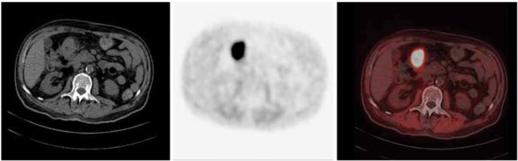
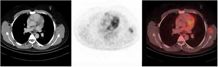
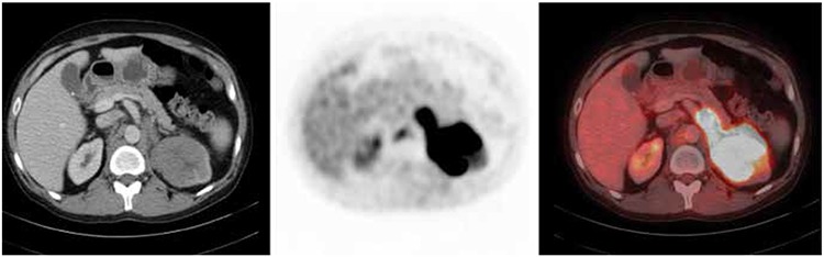
Out of the 82 patients, primary tumor was correctly identified in 57.3% patients by 18F-FDG PET-CT (true positive). The PET-CT scan results were negative for primary site localization in 15% of patients (false negative). While 21% had true negative results, 7.3% displayed false positive results. PET-CT scan upstaged the disease in 27% cases. Overall, the diagnostic accuracy was found to be 78%, sensitivity 80%, specificity 74%, positive predictive value 88.7% and negative predictive value 59%.
Our data supports the utility of 18F-FDG PET-CT scan in the localization and staging of CUP syndrome.
在原发灶不明癌(CUP)综合征患者中检测原发肿瘤部位一直是诊断难题,需要进行广泛检查。早期检测原发肿瘤部位并进行特异性治疗可改善预后。原发肿瘤部位的低检出率可归因于其生物学行为或原发肿瘤体积过小,难以通过传统影像学检测到。本研究的目的是评估18F-氟脱氧葡萄糖(18F-FDG)正电子发射断层扫描-计算机断层扫描(PET-CT)在检测CUP方面的诊断准确性。
对2009年11月至2013年12月期间100例CUP综合征患者的PET-CT扫描进行回顾性横断面分析。18例无法获得最终组织病理学结果以进行相关性分析的患者被排除在分析之外。将高代谢部位与组织病理学进行相关性评估。对PET-CT的诊断准确性、敏感性、特异性、阳性预测值和阴性预测值进行评估。
在82例患者中,18F-FDG PET-CT正确识别出57.3%患者的原发肿瘤(真阳性)。PET-CT扫描结果显示15%患者的原发部位定位为阴性(假阴性)。21%患者为真阴性结果,7.3%患者为假阳性结果。PET-CT扫描使27%的病例分期上升。总体而言,诊断准确性为78%,敏感性为80%,特异性为74%,阳性预测值为88.7%,阴性预测值为59%。
我们的数据支持18F-FDG PET-CT扫描在CUP综合征定位和分期中的应用。