• 文献检索
  • 文档翻译
  • 深度研究
  • 学术资讯
  • Suppr Zotero 插件Zotero 插件
  • 邀请有礼
  • 套餐&价格
  • 历史记录
应用&插件
Suppr Zotero 插件Zotero 插件浏览器插件Mac 客户端Windows 客户端微信小程序
定价
高级版会员购买积分包购买API积分包
服务
文献检索文档翻译深度研究API 文档MCP 服务
关于我们
关于 Suppr公司介绍联系我们用户协议隐私条款
关注我们

Suppr 超能文献

核心技术专利:CN118964589B侵权必究
粤ICP备2023148730 号-1Suppr @ 2026

文献检索

告别复杂PubMed语法,用中文像聊天一样搜索,搜遍4000万医学文献。AI智能推荐,让科研检索更轻松。

立即免费搜索

文件翻译

保留排版,准确专业,支持PDF/Word/PPT等文件格式,支持 12+语言互译。

免费翻译文档

深度研究

AI帮你快速写综述,25分钟生成高质量综述,智能提取关键信息,辅助科研写作。

立即免费体验

正畸诊断中三维面部软组织扫描与头颅侧位X线片的相关性评估

Correlation Assessment between Three-Dimensional Facial Soft Tissue Scan and Lateral Cephalometric Radiography in Orthodontic Diagnosis.

作者信息

Zecca Piero Antonio, Fastuca Rosamaria, Beretta Matteo, Caprioglio Alberto, Macchi Aldo

机构信息

Department of Surgical and Morphological Sciences, University of Insubria, 21100 Varese, Italy.

出版信息

Int J Dent. 2016;2016:1473918. doi: 10.1155/2016/1473918. Epub 2016 May 29.

DOI:10.1155/2016/1473918
PMID:27313615
原文链接:https://pmc.ncbi.nlm.nih.gov/articles/PMC4903122/
Abstract

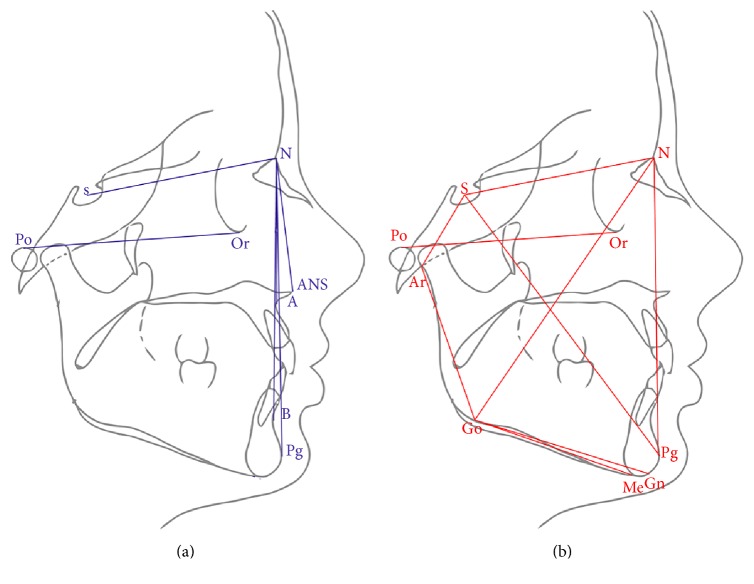
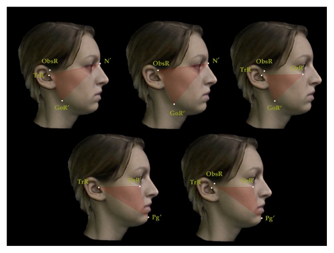
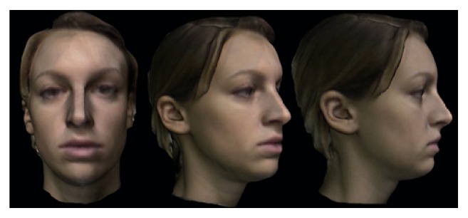
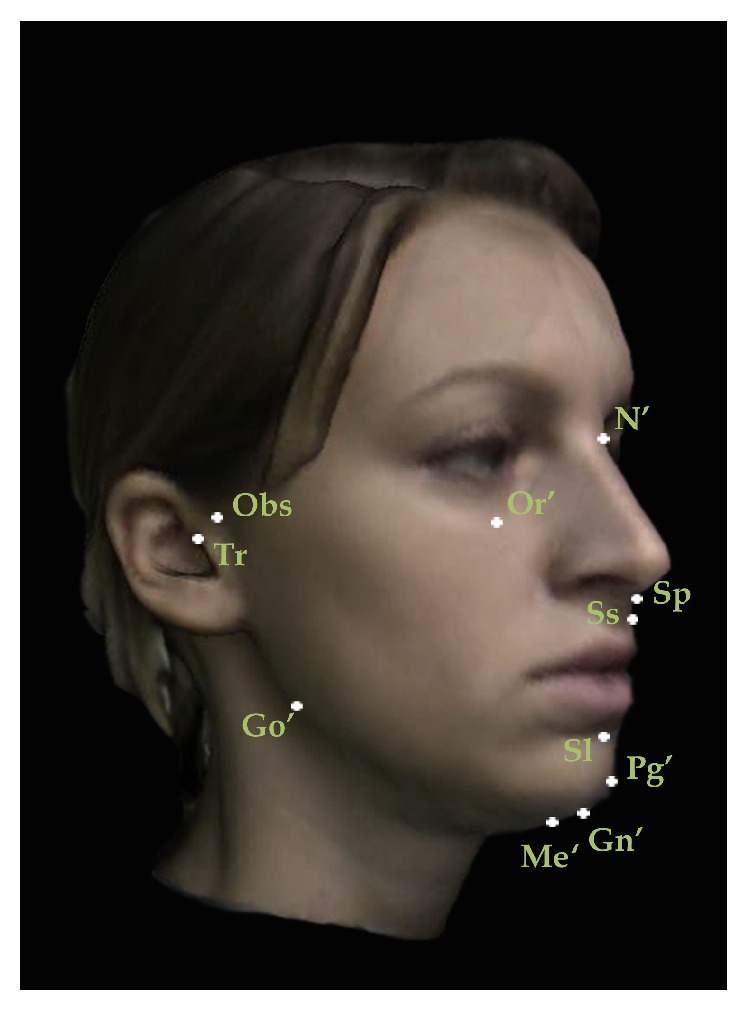
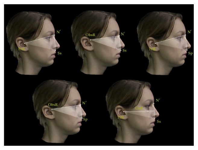
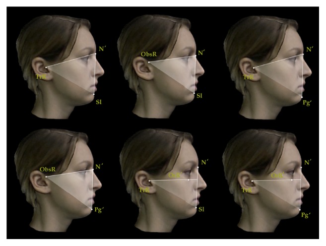
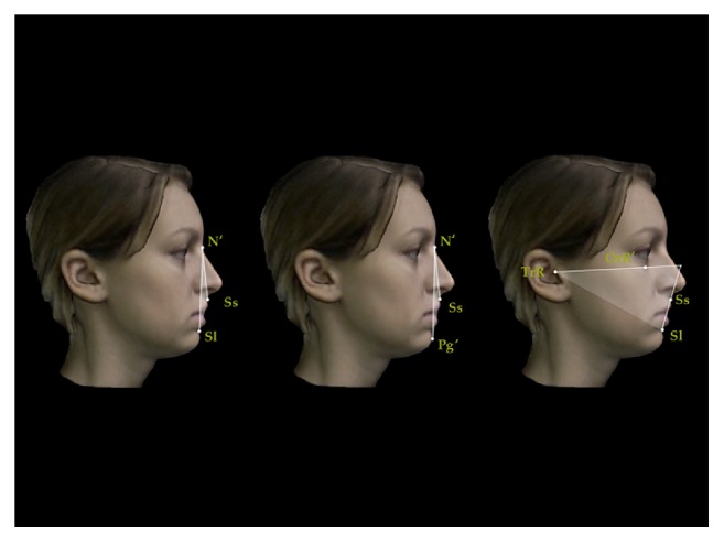
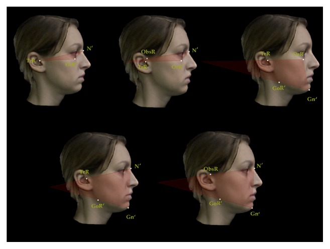
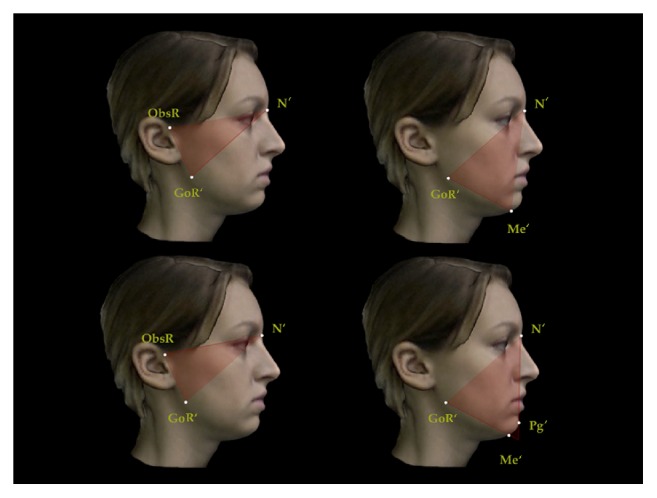
Purpose. The aim of the present prospective study was to investigate correlations between 3D facial soft tissue scan and lateral cephalometric radiography measurements. Materials and Methods. The study sample comprised 312 subjects of Caucasian ethnic origin. Exclusion criteria were all the craniofacial anomalies, noticeable asymmetries, and previous or current orthodontic treatment. A cephalometric analysis was developed employing 11 soft tissue landmarks and 14 sagittal and 14 vertical angular measurements corresponding to skeletal cephalometric variables. Cephalometric analyses on lateral cephalometric radiographies were performed for all subjects. The measurements were analysed in terms of their reliability and gender-age specific differences. Then, the soft tissue values were analysed for any correlations with lateral cephalometric radiography variables using Pearson correlation coefficient analysis. Results. Low, medium, and high correlations were found for sagittal and vertical measurements. Sagittal measurements seemed to be more reliable in providing a soft tissue diagnosis than vertical measurements. Conclusions. Sagittal parameters seemed to be more reliable in providing a soft tissue diagnosis similar to lateral cephalometric radiography. Vertical soft tissue measurements meanwhile showed a little less correlation with the corresponding cephalometric values perhaps due to the low reproducibility of cranial base and mandibular landmarks.

摘要

目的。本前瞻性研究的目的是调查三维面部软组织扫描与头影测量X线侧位片测量之间的相关性。材料与方法。研究样本包括312名高加索人种受试者。排除标准为所有颅面畸形、明显不对称以及既往或当前的正畸治疗。采用11个软组织标志点以及与骨骼头影测量变量对应的14个矢状面和14个垂直角度测量值进行头影测量分析。对所有受试者的头影测量X线侧位片进行头影测量分析。对测量值进行可靠性及性别年龄特异性差异分析。然后,使用Pearson相关系数分析软组织值与头影测量X线侧位片变量之间的相关性。结果。矢状面和垂直测量值存在低、中、高相关性。矢状面测量在提供软组织诊断方面似乎比垂直测量更可靠。结论。矢状面参数在提供类似于头影测量X线侧位片的软组织诊断方面似乎更可靠。同时,垂直软组织测量值与相应的头影测量值的相关性稍低,这可能是由于颅底和下颌标志点的可重复性较低。

https://cdn.ncbi.nlm.nih.gov/pmc/blobs/3ff3/4903122/baea1494fc97/IJD2016-1473918.009.jpg
https://cdn.ncbi.nlm.nih.gov/pmc/blobs/3ff3/4903122/09cadd7d7ff1/IJD2016-1473918.001.jpg
https://cdn.ncbi.nlm.nih.gov/pmc/blobs/3ff3/4903122/444cb5ae1afa/IJD2016-1473918.002.jpg
https://cdn.ncbi.nlm.nih.gov/pmc/blobs/3ff3/4903122/82c5b79470a0/IJD2016-1473918.003.jpg
https://cdn.ncbi.nlm.nih.gov/pmc/blobs/3ff3/4903122/7bea433e4783/IJD2016-1473918.004.jpg
https://cdn.ncbi.nlm.nih.gov/pmc/blobs/3ff3/4903122/6e0646ed92cd/IJD2016-1473918.005.jpg
https://cdn.ncbi.nlm.nih.gov/pmc/blobs/3ff3/4903122/d5298fbf57ed/IJD2016-1473918.006.jpg
https://cdn.ncbi.nlm.nih.gov/pmc/blobs/3ff3/4903122/1daab7751149/IJD2016-1473918.007.jpg
https://cdn.ncbi.nlm.nih.gov/pmc/blobs/3ff3/4903122/e2bbb6cbf9d6/IJD2016-1473918.008.jpg
https://cdn.ncbi.nlm.nih.gov/pmc/blobs/3ff3/4903122/baea1494fc97/IJD2016-1473918.009.jpg

相似文献

1
Correlation Assessment between Three-Dimensional Facial Soft Tissue Scan and Lateral Cephalometric Radiography in Orthodontic Diagnosis.正畸诊断中三维面部软组织扫描与头颅侧位X线片的相关性评估
Int J Dent. 2016;2016:1473918. doi: 10.1155/2016/1473918. Epub 2016 May 29.
2
3D soft tissue analysis--part 2: vertical parameters.三维软组织分析——第2部分:垂直参数
J Orofac Orthop. 2010 May;71(3):207-20. doi: 10.1007/s00056-010-9943-9. Epub 2010 May 26.
3
3D soft tissue analysis--part 1: sagittal parameters.三维软组织分析——第1部分:矢状面参数
J Orofac Orthop. 2010 Jan;71(1):40-52. doi: 10.1007/s00056-010-9926-x. Epub 2010 Feb 5.
4
Facial soft tissue thickness differences among different vertical facial patterns.不同垂直面型的面部软组织厚度差异。
Forensic Sci Int. 2020 Dec;317:110468. doi: 10.1016/j.forsciint.2020.110468. Epub 2020 Sep 9.
5
Comparative study of cephalometric measurements using 3 imaging modalities.三种影像学方法测量头影测量指标的比较研究。
J Am Dent Assoc. 2017 Dec;148(12):913-921. doi: 10.1016/j.adaj.2017.07.030. Epub 2017 Oct 16.
6
New three-dimensional cephalometric analyses among adults with a skeletal Class I pattern and normal occlusion.对具有骨骼I类模式和正常咬合的成年人进行的新的三维头影测量分析。
Korean J Orthod. 2013 Apr;43(2):62-73. doi: 10.4041/kjod.2013.43.2.62. Epub 2013 Apr 25.
7
Skeletal-versus soft-tissue-based cephalometric analyses: is the correlation reproducible?基于骨骼与软组织的头影测量分析:相关性是否具有可重复性?
Acta Odontol Scand. 2019 Mar;77(2):135-141. doi: 10.1080/00016357.2018.1515443. Epub 2018 Nov 3.
8
Photographic Assessment of Cephalometric Measurements in Skeletal Class II Cases: A Comparative Study.骨性II类病例头影测量的摄影评估:一项对比研究。
J Clin Diagn Res. 2017 Jun;11(6):ZC60-ZC64. doi: 10.7860/JCDR/2017/25042.10075. Epub 2017 Jun 1.
9
The Reliability of Three-Dimensional Landmark-Based Craniomaxillofacial and Airway Cephalometric Analysis.基于三维地标点的颅颌面及气道头影测量分析的可靠性
Diagnostics (Basel). 2023 Jul 13;13(14):2360. doi: 10.3390/diagnostics13142360.
10
New Sagittal and Vertical Cephalometric Analysis Methods: A Systematic Review.新的矢状面和垂直头影测量分析方法:系统评价
Diagnostics (Basel). 2022 Jul 15;12(7):1723. doi: 10.3390/diagnostics12071723.

引用本文的文献

1
Innovative Aesthetic and Functional Orthodontic Planning with Hard and Soft Tissue Analyses.通过软硬组织分析进行创新的美学与功能正畸规划。
J Clin Med. 2025 Jun 23;14(13):4458. doi: 10.3390/jcm14134458.
2
Relationship Between Cone-Beam CT Evaluation and Clinical Evaluation Before and After Orthodontic Treatment and the Rate of Gingival Recession: A Systematic Review.正畸治疗前后锥形束CT评估与临床评估的关系及牙龈退缩率:一项系统评价
Cureus. 2024 Jun 17;16(6):e62536. doi: 10.7759/cureus.62536. eCollection 2024 Jun.
3
Correlation Between Cephalometric Values and Soft Tissue Profile in Class I and Class II Adult Patients based on Vertical Patterns.

本文引用的文献

1
Hard and soft tissue correlations in facial profiles: a canonical correlation study.面部轮廓中软硬组织的相关性:一项典型相关性研究。
Clin Cosmet Investig Dent. 2015 Jan 12;7:9-15. doi: 10.2147/CCIDE.S73457. eCollection 2015.
2
How important are lateral cephalometric radiographs in orthodontic treatment planning?侧颅面 X 光片在正畸治疗计划中有多重要?
Am J Orthod Dentofacial Orthop. 2011 Feb;139(2):e175-81. doi: 10.1016/j.ajodo.2010.09.021.
3
3D soft tissue analysis--part 2: vertical parameters.三维软组织分析——第2部分:垂直参数
基于垂直模式的I类和II类成年患者头影测量值与软组织侧貌的相关性
Turk J Orthod. 2024 Mar 28;37(1):36-43. doi: 10.4274/TurkJOrthod.2023.2022.20.
4
Efficacy of the combination of myofunctional therapy (lip exercises) and activator high-pull headgear in the closure of interlabial gap in long-face skeletal class II patients with lip incompetence: A 6-8-month longitudinal randomized clinical trial.肌功能治疗(唇部训练)与Activator高位牵引矫治器联合应用对长面型骨性Ⅱ类唇功能不全患者唇间隙关闭的疗效:一项6 - 8个月的纵向随机临床试验。
Dent Res J (Isfahan). 2024 Jan 25;21:3. eCollection 2024.
5
The Reliability of Three-Dimensional Landmark-Based Craniomaxillofacial and Airway Cephalometric Analysis.基于三维地标点的颅颌面及气道头影测量分析的可靠性
Diagnostics (Basel). 2023 Jul 13;13(14):2360. doi: 10.3390/diagnostics13142360.
6
Treatment outcome of class II malocclusion therapy including extraction of maxillary first molars: a cephalometric comparison between normodivergent and hyperdivergent facial types.上颌第一磨牙拔除的安氏Ⅱ类错(牙合)矫治的治疗效果:正常生长型与高角型颅面结构的头影测量比较。
PeerJ. 2022 Dec 12;10:e14537. doi: 10.7717/peerj.14537. eCollection 2022.
7
Skeletal, Dental and Soft Tissue Cephalometric Changes after Orthodontic Treatment of Dental Class II Malocclusion with Maxillary First Molar or First Premolar Extractions.拔除上颌第一磨牙或第一前磨牙矫治安氏II类错牙合畸形后的骨骼、牙齿及软组织头影测量变化
J Clin Med. 2022 Jun 2;11(11):3170. doi: 10.3390/jcm11113170.
8
Reliability of Photogrammetric Landmarks to the Conventional Cephalogram for Analyzing Soft-Tissue Landmarks in Orthodontics.用于正畸学中分析软组织标志点的摄影测量标志点相对于传统头影测量的可靠性
J Pharm Bioallied Sci. 2021 Jun;13(Suppl 1):S171-S175. doi: 10.4103/jpbs.JPBS_634_20. Epub 2021 Jun 5.
9
Influence of facial components in class III malocclusion esthetic perception of orthodontists, patients, and laypersons.正畸医生、患者和非专业人士对面部特征在 III 类错牙合畸形美观感知中的影响。
J Orofac Orthop. 2022 Jan;83(1):48-58. doi: 10.1007/s00056-021-00287-1. Epub 2021 Apr 30.
10
Midpalatal Suture Density Evaluation after Rapid and Slow Maxillary Expansion with a Low-Dose CT Protocol: A Retrospective Study.低剂量 CT 方案评估快速与慢速上颌扩张后正中缝密度:一项回顾性研究。
Medicina (Kaunas). 2020 Mar 5;56(3):112. doi: 10.3390/medicina56030112.
J Orofac Orthop. 2010 May;71(3):207-20. doi: 10.1007/s00056-010-9943-9. Epub 2010 May 26.
4
3D soft tissue analysis--part 1: sagittal parameters.三维软组织分析——第1部分:矢状面参数
J Orofac Orthop. 2010 Jan;71(1):40-52. doi: 10.1007/s00056-010-9926-x. Epub 2010 Feb 5.
5
Soft tissue profile changes following maxillary protraction in Class III subjects.上颌骨前牵引后 III 类错(牙合)患者软组织侧貌变化。
Eur J Orthod. 2010 Aug;32(4):419-24. doi: 10.1093/ejo/cjp119. Epub 2010 Jan 6.
6
Soft tissue images from cephalograms compared with those from a 3D surface acquisition system.头影测量片的软组织图像与三维表面采集系统的图像比较。
Angle Orthod. 2010 Jan;80(1):58-64. doi: 10.2319/111408-583.1.
7
Three-dimensional analysis of facial morphology surface changes in untreated children from 12 to 14 years of age.12至14岁未接受治疗儿童面部形态表面变化的三维分析。
Am J Orthod Dentofacial Orthop. 2008 Dec;134(6):751-60. doi: 10.1016/j.ajodo.2007.01.037.
8
The influence of cephalometrics on orthodontic treatment planning.头影测量学对正畸治疗计划的影响。
Eur J Orthod. 2008 Dec;30(6):630-5. doi: 10.1093/ejo/cjn059. Epub 2008 Nov 3.
9
Three-dimensional analysis of changes of the malar-midfacial region after LeFort I osteotomy and maxillary advancement.LeFort I型截骨术及上颌前徙术后颧-面中部区域变化的三维分析
Oral Maxillofac Surg. 2008 May;12(1):5-12. doi: 10.1007/s10006-008-0094-8.
10
The effect of sex and age on facial asymmetry in healthy subjects: a cross-sectional study from adolescence to mid-adulthood.性别和年龄对健康受试者面部不对称的影响:一项从青春期到中年期的横断面研究。
J Oral Maxillofac Surg. 2001 Apr;59(4):382-8. doi: 10.1053/joms.2001.21872.