Lu Sheng-Feng, Huang Yan, Wang Ning, Shen Wei-Xing, Fu Shu-Ping, Li Qian, Yu Mei-Ling, Liu Wan-Xin, Chen Xia, Jing Xin-Yue, Zhu Bing-Mei
Key Laboratory of Acupuncture and Medicine Research of Ministry of Education, Nanjing University of Chinese Medicine, Nanjing 210023, China.
Key Laboratory of Acupuncture and Medicine Research of Ministry of Education, Nanjing University of Chinese Medicine, Nanjing 210023, China; Key Laboratory of Acupuncture and Immunological Effects, Shanghai University of Traditional Chinese Medicine, Shanghai 200030, China.
Evid Based Complement Alternat Med. 2016;2016:4609784. doi: 10.1155/2016/4609784. Epub 2016 May 25.
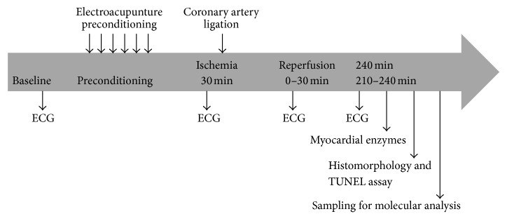
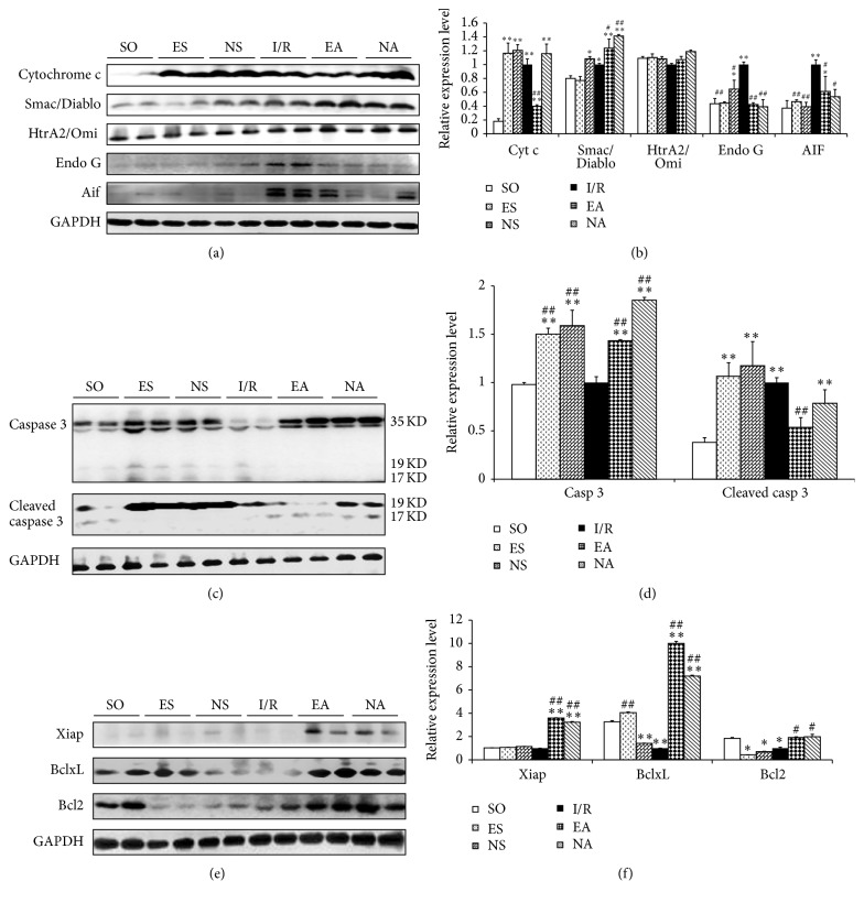
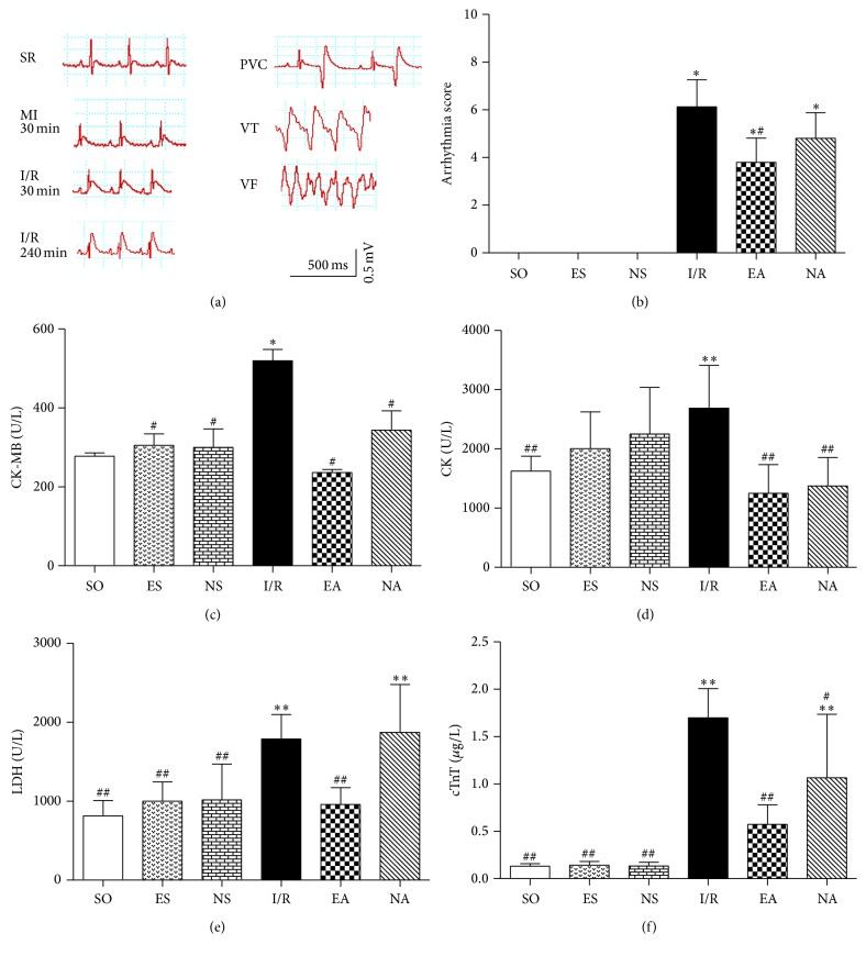
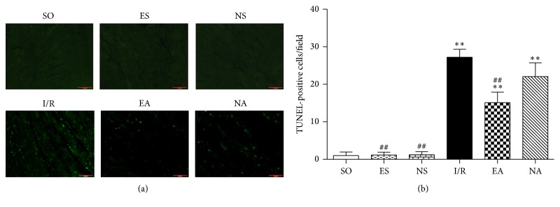
Objectives. Our previous study has used RNA-seq technology to show that apoptotic molecules were involved in the myocardial protection of electroacupuncture pretreatment (EAP) on the ischemia/reperfusion (I/R) animal model. Therefore, this study was designed to investigate how EAP protects myocardium against myocardial I/R injury through antiapoptotic mechanism. Methods. By using rats with myocardial I/R, we ligated the left anterior descending artery (LAD) for 30 minutes followed by 4 hr of reperfusion after EAP at the Neiguan (PC6) acupoint for 12 days; we employed arrhythmia scores, serum myocardial enzymes, and cardiac troponin T (cTnT) to evaluate the cardioprotective effect. Heart tissues were harvested for western blot analyses for the expressions of pro- and antiapoptotic signaling molecules. Results. Our preliminary findings showed that EAP increased the survival of the animals along with declined arrhythmia scores and decreased CK, LDH, CK-Mb, and cTnT levels. Further analyses with the heart tissues detected reduced myocardial fiber damage, decreased number of apoptotic cells and the protein expressions of Cyt c and cleaved caspase 3, and the elevated level of Endo G and AIF after EAP intervention. At the same time, the protein expressions of antiapoptotic molecules, including Xiap, BclxL, and Bcl2, were obviously increased. Conclusions. The present study suggested that EAP protected the myocardium from I/R injury at least partially through the activation of endogenous antiapoptotic signaling.
目的。我们之前的研究使用RNA测序技术表明,凋亡分子参与了电针预处理(EAP)对缺血/再灌注(I/R)动物模型的心肌保护作用。因此,本研究旨在探讨EAP如何通过抗凋亡机制保护心肌免受心肌I/R损伤。方法。我们使用心肌I/R大鼠,在左侧内关穴(PC6)进行12天的EAP后,结扎左前降支动脉(LAD)30分钟,随后再灌注4小时;我们采用心律失常评分、血清心肌酶和心肌肌钙蛋白T(cTnT)来评估心脏保护作用。采集心脏组织进行蛋白质印迹分析,以检测促凋亡和抗凋亡信号分子的表达。结果。我们的初步研究结果表明,EAP提高了动物的存活率,同时降低了心律失常评分,并降低了CK、LDH、CK-Mb和cTnT水平。对心脏组织的进一步分析发现,EAP干预后心肌纤维损伤减轻,凋亡细胞数量减少,Cyt c和裂解的caspase 3的蛋白质表达降低,Endo G和AIF水平升高。同时,包括Xiap、BclxL和Bcl2在内的抗凋亡分子的蛋白质表达明显增加。结论。本研究表明,EAP至少部分通过激活内源性抗凋亡信号来保护心肌免受I/R损伤。