Serra Laura, Bianchi Guendalina, Bruschini Michela, Giulietti Giovanni, Domenico Carlotta Di, Bonarota Sabrina, Petrucci Antonio, Silvestri Gabriella, Perna Alessia, Meola Giovanni, Caltagirone Carlo, Bozzali Marco
Neuroimaging Laboratory, IRCCS Fondazione Santa Lucia, Rome, Italy.
UOC Neurologia e Neurofisiopatologia, AO San Camillo Forlanini, Rome, Italy.
Front Neurol. 2020 Feb 28;11:113. doi: 10.3389/fneur.2020.00113. eCollection 2020.
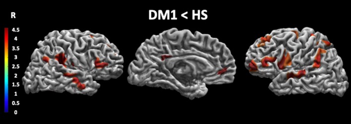
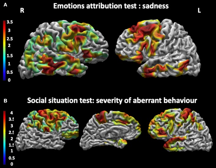
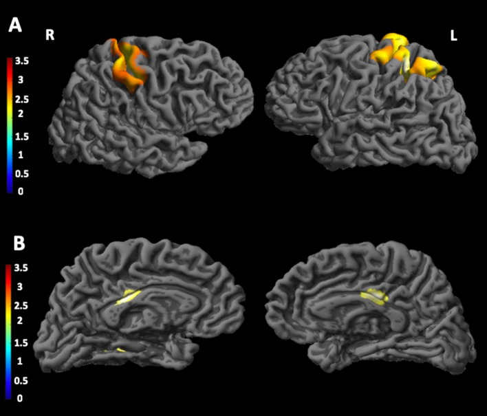
To investigate the cortical thickness in myotonic dystrophy type 1 (DM1) and its potential association with patients' genetic triplet expansion and social cognition deficits. Thirty patients with DM1 underwent the Social Cognition Battery Test and magnetic resonance imaging (MRI) scanning at 3 T. Twenty-five healthy subjects (HSs) were enrolled in the study to serve as a control group for structural MRI data. To assess changes in cortical thickness in DM1 patients, they were compared to HSs using a -test model. Correlations were used to assess potential associations between genetic and clinical characteristics and social cognition performances in the patient group. Additionally, multiple regression models were used to explore associations between cortical thickness, CTG triplet expansion size, and scores obtained by DM1 patients on the Social Cognition Battery. DM1 patients showed low performances in several subtests of the Social Cognition Battery. Specifically, they obtained pathological scores at Emotion Attribution Test (i.e., Sadness, Embarrassment, Happiness, and Anger) and at the Social Situations Test (i.e., recognition of normal situation, recognition of aberrant behavior). Significant negative correlations were found between CTG triplet expansion size and Embarrassment, and Severity of Aberrant Behavior. Similarly, a negative correlation was found between patients' MIRS scores and Sadness. DM1 patients compared to HSs showed reduced thickness in the right premotor cortex, angular gyrus, precuneus, and inferior parietal lobule. Significant associations were found between patients' CTG triplet expansion size and thickness in left postcentral gyrus and in the left primary somatosensory cortex, in the posterior cingulate cortex bilaterally, and in the right lingual gyrus. Finally, significant associations were found between cortical thickness and sadness in the superior temporal gyrus, the right precentral gyrus, the right angular gyrus, and the left medial frontal gyrus bilaterally. DM1 patients showed a negative correlation between cortical thickness in the bilateral precuneus and in the left lateral occipital cortex and performance at the Social Situations Test. Finally, DM1 patients showed a negative correlation between cortical thickness in the left precuneus and in the superior frontal gyrus and scores at the Moral Distinction Test. The present study shows both cortical thickness changes in DM1 patients compared to controls and significant associations between cortical thickness and patients' social cognition performances. These data confirm the presence of widespread brain damages associated with cognitive impairment in DM1 patients.
为研究1型强直性肌营养不良(DM1)患者的皮质厚度及其与患者基因三联体扩增和社会认知缺陷的潜在关联。30例DM1患者接受了社会认知电池测试和3T磁共振成像(MRI)扫描。25名健康受试者(HSs)参与研究,作为结构MRI数据的对照组。为评估DM1患者皮质厚度的变化,采用t检验模型将他们与HSs进行比较。相关性分析用于评估患者组中基因和临床特征与社会认知表现之间的潜在关联。此外,多元回归模型用于探索皮质厚度、CTG三联体扩增大小与DM1患者在社会认知电池测试中获得的分数之间的关联。DM1患者在社会认知电池测试的几个子测试中表现较差。具体而言,他们在情感归因测试(即悲伤、尴尬、快乐和愤怒)以及社会情境测试(即正常情境识别、异常行为识别)中获得了病理分数。CTG三联体扩增大小与尴尬以及异常行为严重程度之间存在显著负相关。同样,患者的MIRS分数与悲伤之间也存在负相关。与HSs相比,DM1患者右侧运动前皮质、角回、楔前叶和顶下小叶厚度减小。患者的CTG三联体扩增大小与左侧中央后回、左侧初级体感皮层、双侧后扣带回皮层以及右侧舌回的厚度之间存在显著关联。最后,在颞上回、右侧中央前回、右侧角回和双侧左侧内侧额叶回中,皮质厚度与悲伤之间存在显著关联。DM1患者双侧楔前叶和左侧枕外侧皮质的皮质厚度与社会情境测试表现之间呈负相关。最后,DM1患者左侧楔前叶和额上回的皮质厚度与道德辨别测试分数之间呈负相关。本研究表明,与对照组相比,DM1患者存在皮质厚度变化,且皮质厚度与患者的社会认知表现之间存在显著关联。这些数据证实了DM1患者存在与认知障碍相关的广泛脑损伤。