Hänggi Jürgen, Bellwald Dorian, Brugger Peter
Division Neuropsychology, Department of Psychology, University of Zurich, Switzerland.
Division Neuropsychology, Department of Psychology, University of Zurich, Switzerland.
Neuroimage Clin. 2016 May 30;11:760-769. doi: 10.1016/j.nicl.2016.05.015. eCollection 2016.
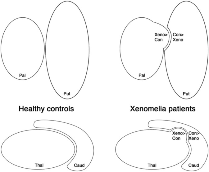
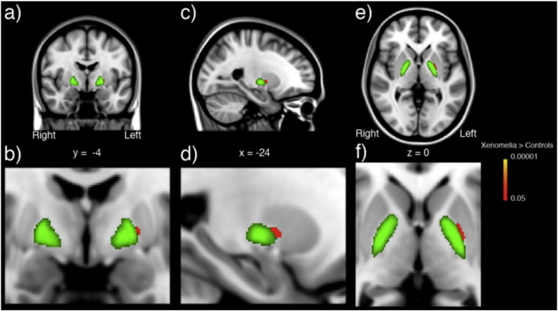
Xenomelia is a rare condition characterized by the persistent desire for the amputation of physically healthy limbs. Associations with morphological alterations such as reduced cortical thickness and surface area. Nothing is known, however, about the potential involvement of subcortical structures. The thalamus and basal ganglia process, relay, and integrate sensorimotor information and are involved in the preparation and execution of movements. Moreover, both of these structures house somatotopic representations of all body parts. We therefore investigated subcortical correlates of xenomelia by assessing basal ganglia and thalamus by means of vertex-wise shape analyses. For that purpose, we compared the shape of the thalamus, putamen, caudate nucleus, and the pallidum in 13 men suffering from xenomelia, all desiring a leg amputation, compared to 13 healthy control men. We hypothesised that the target leg is misrepresented in subcortical structures of individuals with xenomelia, especially in locations with a somatotopic representation. Shape analyses showed thinning of bilateral dorsomedial putamina, left ventromedial caudate nucleus and left medial pallidum associated with xenomelia. This was accompanied by thickening of bilateral lateral pallida and the left frontolateral thalamus. These shape differences were mainly located in sensorimotor areas of somatotopic leg representations. The present study provides strong evidence for shape differences in striatal, pallidal, and thalamic subregions housing subcortical body part representations. It adds to previously described neural correlates of a condition one can barely empathize with and invites future connectivity analyses in cortico-subcortical networks.
肢体完整性认同障碍是一种罕见病症,其特征是持续渴望截去身体健全的肢体。该病症与诸如皮质厚度和表面积减少等形态学改变有关。然而,关于皮质下结构的潜在参与情况却一无所知。丘脑和基底神经节处理、传递并整合感觉运动信息,且参与运动的准备和执行。此外,这两个结构都存在所有身体部位的躯体定位表征。因此,我们通过顶点形状分析评估基底神经节和丘脑,来研究肢体完整性认同障碍的皮质下相关因素。为此,我们将13名患有肢体完整性认同障碍且都渴望截肢的男性的丘脑、壳核、尾状核和苍白球的形状,与13名健康对照男性进行了比较。我们假设,在患有肢体完整性认同障碍的个体的皮质下结构中,尤其是在具有躯体定位表征的位置,目标腿的表征存在错误。形状分析显示,与肢体完整性认同障碍相关的是双侧背内侧壳核、左侧腹内侧尾状核和左侧内侧苍白球变薄。同时伴有双侧外侧苍白球和左侧额外侧丘脑增厚。这些形状差异主要位于躯体定位腿部表征的感觉运动区域。本研究为包含皮质下身体部位表征的纹状体、苍白球和丘脑亚区域的形状差异提供了有力证据。它补充了之前所描述的一种几乎无法让人感同身受的病症的神经相关性,并促使未来对皮质 - 皮质下网络进行连通性分析。