Beqollari Donald, Romberg Christin F, Dobrowolny Gabriella, Martini Martina, Voss Andrew A, Musarò Antonio, Bannister Roger A
Department of Medicine-Cardiology Division, University of Colorado School of Medicine, 12700 East 19th Avenue, B-139, Aurora, CO 80045 USA.
Institute Pasteur Cenci-Bolognetti, DAHFMO-Unit of Histology and Medical Embryology, La Sapienza University, Via A. Scarpa, 14, 00161 Rome, Italy ; Center for Life Nano Science@Sapienza, Istituto Italiano di Tecnologia, Rome, Italy.
Skelet Muscle. 2016 Jun 23;6:24. doi: 10.1186/s13395-016-0094-6. eCollection 2016.
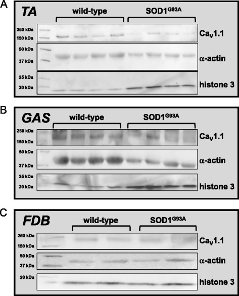
Amyotrophic lateral sclerosis (ALS) is an adult-onset neurodegenerative disorder that is typically fatal within 3-5 years of diagnosis. While motoneuron death is the defining characteristic of ALS, the events that underlie its pathology are not restricted to the nervous system. In this regard, ALS muscle atrophies and weakens significantly before presentation of neurological symptoms. Since the skeletal muscle L-type Ca(2+) channel (CaV1.1) is a key regulator of both mass and force, we investigated whether CaV1.1 function is impaired in the muscle of two distinct mouse models carrying an ALS-linked mutation.
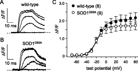
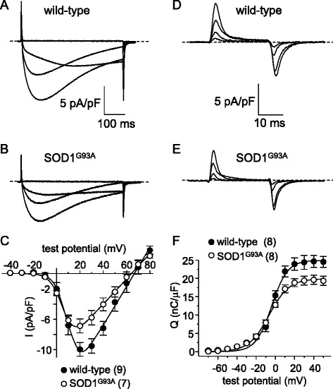
We recorded L-type currents, charge movements, and myoplasmic Ca(2+) transients from dissociated flexor digitorum brevis (FDB) fibers to assess CaV1.1 function in two mouse models expressing a type 1 Cu/Zn superoxide dismutase mutant (SOD1(G93A)).
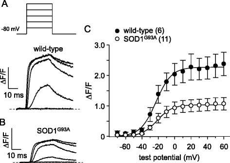
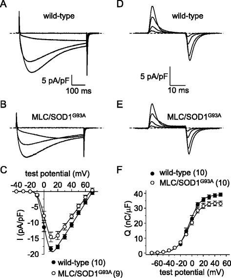
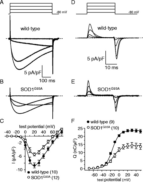
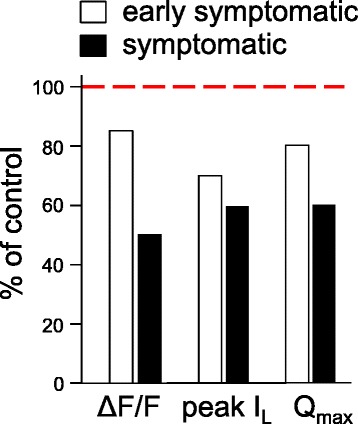
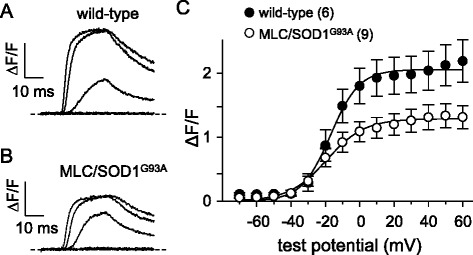
In FDB fibers obtained from "symptomatic" global SOD1(G93A) mice, we observed a substantial reduction of SR Ca(2+) release in response to depolarization relative to fibers harvested from age-matched control mice. L-type current and charge movement were both reduced by 40 % in symptomatic SOD1(G93A) fibers when compared to control fibers. Ca(2+) transients were not significantly reduced in similar experiments performed with FDB fibers obtained from "early-symptomatic" SOD1(G93A) mice, but L-type current and charge movement were decreased (30 and 20 %, respectively). Reductions in SR Ca(2+) release (35 %), L-type current (20 %), and charge movement (15 %) were also observed in fibers obtained from another model where SOD1(G93A) expression was restricted to skeletal muscle.
We report reductions in EC coupling, L-type current density, and charge movement in FDB fibers obtained from symptomatic global SOD1(G93A) mice. Experiments performed with FDB fibers obtained from early-symptomatic SOD1(G93A) and skeletal muscle autonomous MLC/SOD1(G93A) mice support the idea that events occurring locally in the skeletal muscle contribute to the impairment of CaV1.1 function in ALS muscle independently of innervation status.
肌萎缩侧索硬化症(ALS)是一种成年发病的神经退行性疾病,通常在确诊后3至5年内致命。虽然运动神经元死亡是ALS的决定性特征,但其病理过程所涉及的事件并不局限于神经系统。在这方面,ALS患者在出现神经症状之前,肌肉就会显著萎缩和无力。由于骨骼肌L型钙通道(CaV1.1)是肌肉质量和力量的关键调节因子,我们研究了携带ALS相关突变的两种不同小鼠模型的肌肉中CaV1.1功能是否受损。
我们从解离的趾短屈肌(FDB)纤维记录L型电流、电荷移动和肌浆钙瞬变,以评估两种表达1型铜/锌超氧化物歧化酶突变体(SOD1(G93A))的小鼠模型中CaV1.1的功能。
在从“有症状的”全身性SOD1(G93A)小鼠获得的FDB纤维中,相对于从年龄匹配的对照小鼠收获的纤维,我们观察到去极化后肌浆网钙释放大幅减少。与对照纤维相比,有症状的SOD1(G93A)纤维中的L型电流和电荷移动均减少了约40%。在用从“早期有症状的”SOD1(G93A)小鼠获得的FDB纤维进行的类似实验中,钙瞬变没有显著减少,但L型电流和电荷移动减少了(分别约为30%和20%)。在从另一种将SOD1(G93A)表达限制在骨骼肌的模型获得的纤维中,也观察到肌浆网钙释放减少(约35%)、L型电流减少(约20%)和电荷移动减少(约15%)。
我们报告了从有症状的全身性SOD1(G93A)小鼠获得的FDB纤维中兴奋-收缩偶联、L型电流密度和电荷移动减少。用从早期有症状的SOD1(G93A)和骨骼肌自主性MLC/SOD1(G93A)小鼠获得的FDB纤维进行的实验支持这样一种观点,即骨骼肌局部发生的事件导致ALS肌肉中CaV1.1功能受损,而与神经支配状态无关。