Surova Yulia, Lampinen Björn, Nilsson Markus, Lätt Jimmy, Hall Sara, Widner Håkan, van Westen Danielle, Hansson Oskar
Department of Clinical Sciences, Lund, Lund University, Lund, Sweden.
Department of Neurology, Skåne University Hospital, Lund, Sweden.
PLoS One. 2016 Jun 30;11(6):e0157755. doi: 10.1371/journal.pone.0157755. eCollection 2016.
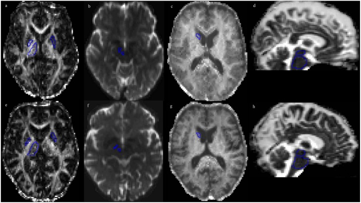
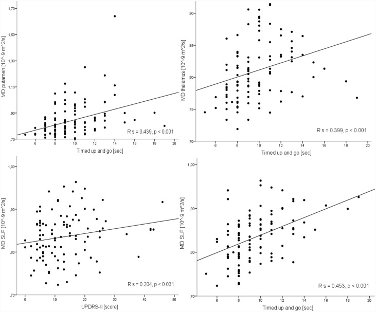
In Parkinson's disease (PD), pathological microstructural changes occur and such changes might be detected using diffusion magnetic resonance imaging (dMRI). However, it is unclear whether dMRI improves PD diagnosis or helps differentiating between phenotypes, such as postural instability gait difficulty (PIGD) and tremor dominant (TD) PD. We included 105 patients with PD and 44 healthy controls (HC), all of whom underwent dMRI as part of the prospective Swedish BioFINDER study. Diffusion kurtosis imaging (DKI) and neurite density imaging (NDI) analyses were performed using regions of interest in the basal ganglia, the thalamus, the pons and the midbrain as well as tractography of selected white matter tracts. In the putamen, the PD group showed increased mean diffusivity (MD) (p = .003), decreased fractional anisotropy (FA) (p = .001) and decreased mean kurtosis (MK), compared to HC (p = .024). High MD and a low MK in the putamen were associated with more severe motor and cognitive symptomatology (p < .05). Also, patients with PIGD exhibited increased MD in the putamen compared to the TD patients (p = .009). In the thalamus, MD was increased (p = .001) and FA was decreased (p = .032) in PD compared to HC. Increased MD and decreased FA correlated negatively with motor speed and balance (p < .05). In the superior longitudinal fasciculus (SLF), MD (p = .019) and fiso were increased in PD compared to HC (p = .03). These changes correlated negatively with motor speed (p < .002) and balance (p < .037). However, most of the observed changes in PD were also present in cases with either multiple system atrophy (n = 11) or progressive supranuclear palsy (n = 10). In conclusion, PD patients exhibit microstructural changes in the putamen, the thalamus, and the SLF, which are associated with worse disease severity. However, the dMRI changes are not sufficiently specific to improve the diagnostic work-up of PD. Longitudinal studies should evaluate whether dMRI measures can be used to track disease progression.
在帕金森病(PD)中,会出现病理性微观结构变化,且这些变化或许可通过扩散磁共振成像(dMRI)检测到。然而,目前尚不清楚dMRI是否能改善PD的诊断,或有助于区分不同的表型,如姿势不稳步态障碍(PIGD)型和震颤为主(TD)型PD。我们纳入了105例PD患者和44名健康对照者(HC),他们均接受了dMRI检查,这是前瞻性瑞典生物探索者研究的一部分。使用基底神经节、丘脑、脑桥和中脑的感兴趣区域进行扩散峰度成像(DKI)和神经突密度成像(NDI)分析,以及对选定白质束进行纤维束成像。与HC相比,PD组壳核的平均扩散率(MD)增加(p = 0.003),分数各向异性(FA)降低(p = 0.001),平均峰度(MK)降低(p = 0.024)。壳核中高MD和低MK与更严重的运动和认知症状相关(p < 0.05)。此外,与TD患者相比,PIGD患者壳核的MD增加(p = 0.009)。与HC相比,PD患者丘脑的MD增加(p = 0.001),FA降低(p = 0.032)。MD增加和FA降低与运动速度和平衡呈负相关(p < 0.05)。与HC相比,PD患者的上纵束(SLF)中MD(p = 0.019)和fiso增加(p = 0.03)。这些变化与运动速度(p < 0.002)和平衡(p < 0.037)呈负相关。然而,在多系统萎缩(n = 11)或进行性核上性麻痹(n = 10)患者中也出现了PD中观察到的大多数变化。总之,PD患者在壳核、丘脑和SLF中表现出微观结构变化,这些变化与更严重的疾病严重程度相关。然而,dMRI变化特异性不足,无法改善PD的诊断检查。纵向研究应评估dMRI测量是否可用于跟踪疾病进展。