Huang Ya-Qin, Liang He-Yue, Yang Zhao-Xia, Ding Ying, Zeng Meng-Su, Rao Sheng-Xiang
aDepartment of Radiology, Zhongshan Hospital, Fudan University, Shanghai, China bShanghai Medical Imaging Institute, Shanghai, China cDepartment of Radiology, The Ningbo First Hospital, Ningbo, China.
Medicine (Baltimore). 2016 Jun;95(26):e4034. doi: 10.1097/MD.0000000000004034.
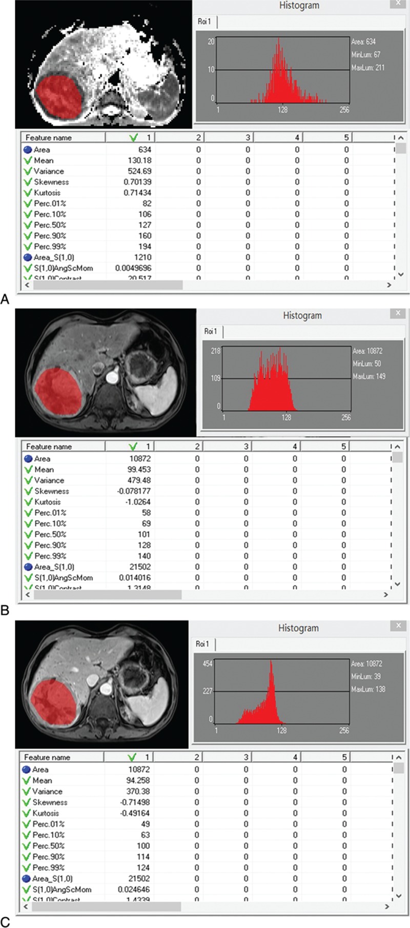
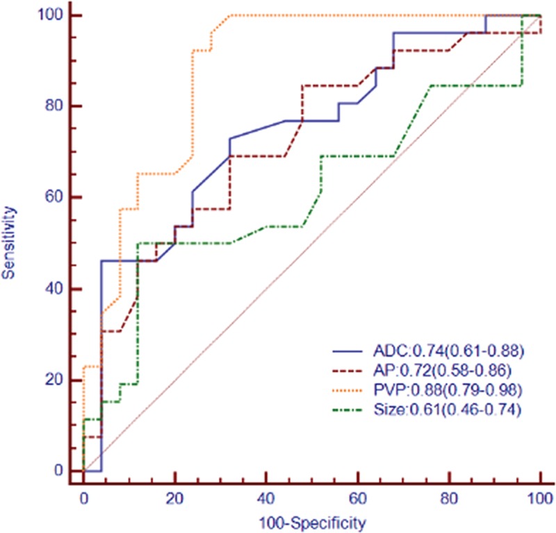
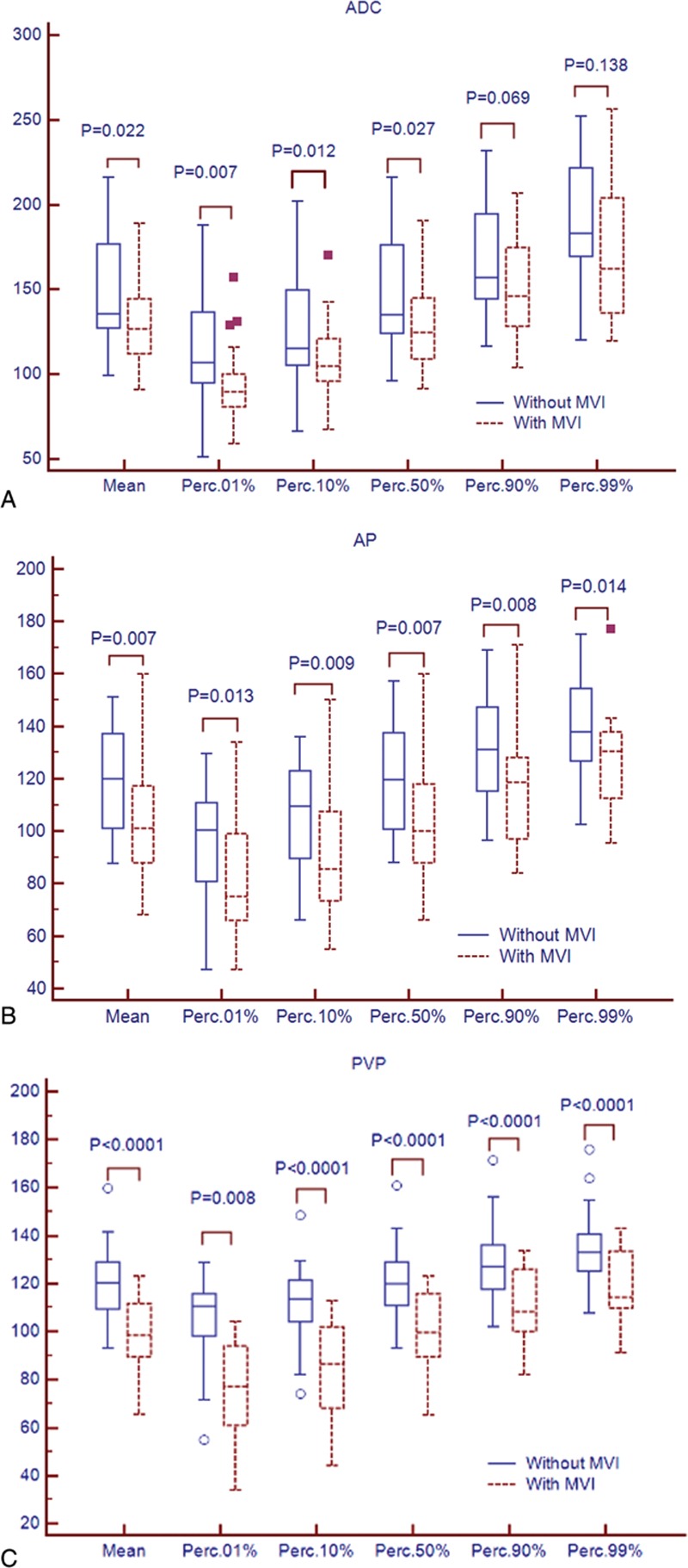
The objective is to explore the value of preoperative magnetic resonance (MR) histogram analyses in predicting microvascular invasion (MVI) of hepatocellular carcinoma (HCC).Fifty-one patients with histologically confirmed HCC who underwent diffusion-weighted and contrast-enhanced MR imaging were included. Histogram analyses were performed and mean, variance, skewness, kurtosis, 1th, 10th, 50th, 90th, and 99th percentiles were derived. Quantitative histogram parameters were compared between HCCs with and without MVI. Receiver operating characteristics (ROC) analyses were generated to compare the diagnostic performance of tumor size, histogram analyses of apparent diffusion coefficient (ADC) maps, and MR enhancement.The mean, 1th, 10th, and 50th percentiles of ADC maps, and the mean, variance. 1th, 10th, 50th, 90th, and 99th percentiles of the portal venous phase (PVP) images were significantly different between the groups with and without MVI (P <0.05), with area under the ROC curves (AUCs) of 0.66 to 0.74 for ADC and 0.76 to 0.88 for PVP. The largest AUC of PVP (1th percentile) showed significantly higher accuracy compared with that of arterial phase (AP) or tumor size (P <0.001).MR histogram analyses-in particular for 1th percentile for PVP images-held promise for prediction of MVI of HCC.
目的是探讨术前磁共振(MR)直方图分析在预测肝细胞癌(HCC)微血管侵犯(MVI)中的价值。纳入了51例经组织学证实为HCC且接受了扩散加权和对比增强MR成像的患者。进行了直方图分析,并得出了平均值、方差、偏度、峰度、第1百分位数、第10百分位数、第50百分位数、第90百分位数和第99百分位数。比较了有和无MVI的HCC之间的定量直方图参数。生成了受试者操作特征(ROC)分析,以比较肿瘤大小、表观扩散系数(ADC)图的直方图分析和MR增强的诊断性能。有和无MVI的组之间,ADC图的平均值、第1百分位数、第10百分位数和第50百分位数,以及门静脉期(PVP)图像的平均值、方差、第1百分位数、第10百分位数、第50百分位数、第90百分位数和第99百分位数存在显著差异(P<0.05),ADC的ROC曲线下面积(AUC)为0.66至0.74,PVP为0.76至0.88。PVP(第1百分位数)的最大AUC显示出与动脉期(AP)或肿瘤大小相比显著更高的准确性(P<0.001)。MR直方图分析——特别是PVP图像的第1百分位数——有望用于预测HCC的MVI。