• 文献检索
  • 文档翻译
  • 深度研究
  • 学术资讯
  • Suppr Zotero 插件Zotero 插件
  • 邀请有礼
  • 套餐&价格
  • 历史记录
应用&插件
Suppr Zotero 插件Zotero 插件浏览器插件Mac 客户端Windows 客户端微信小程序
定价
高级版会员购买积分包购买API积分包
服务
文献检索文档翻译深度研究API 文档MCP 服务
关于我们
关于 Suppr公司介绍联系我们用户协议隐私条款
关注我们

Suppr 超能文献

核心技术专利:CN118964589B侵权必究
粤ICP备2023148730 号-1Suppr @ 2026

文献检索

告别复杂PubMed语法,用中文像聊天一样搜索,搜遍4000万医学文献。AI智能推荐,让科研检索更轻松。

立即免费搜索

文件翻译

保留排版,准确专业,支持PDF/Word/PPT等文件格式,支持 12+语言互译。

免费翻译文档

深度研究

AI帮你快速写综述,25分钟生成高质量综述,智能提取关键信息,辅助科研写作。

立即免费体验

扩散峰度成像和体素内不相干运动扩散加权成像参数在肝细胞癌病理分级和微血管侵犯术前评估中的作用

The Roles of Diffusion Kurtosis Imaging and Intravoxel Incoherent Motion Diffusion-Weighted Imaging Parameters in Preoperative Evaluation of Pathological Grades and Microvascular Invasion in Hepatocellular Carcinoma.

作者信息

Wang Fei, Yan Chun Yue, Wang Cai Hong, Yang Yan, Zhang Dong

机构信息

Department of Medical Imaging, Luzhou People's Hospital, Luzhou, China.

Department of Radiology, Xinqiao Hospital, Third Military Medical University, Chongqing, China.

出版信息

Front Oncol. 2022 May 11;12:884854. doi: 10.3389/fonc.2022.884854. eCollection 2022.

DOI:10.3389/fonc.2022.884854
PMID:35646649
原文链接:https://pmc.ncbi.nlm.nih.gov/articles/PMC9131658/
Abstract

BACKGROUND

Currently, there are disputes about the parameters of diffusion kurtosis imaging (DKI), intravoxel incoherent motion (IVIM), and diffusion-weighted imaging (DWI) in predicting pathological grades and microvascular invasion (MVI) in hepatocellular carcinoma (HCC). The aim of our study was to investigate and compare the predictive power of DKI and IVIM-DWI parameters for preoperative evaluation of pathological grades and MVI in HCC.

METHODS

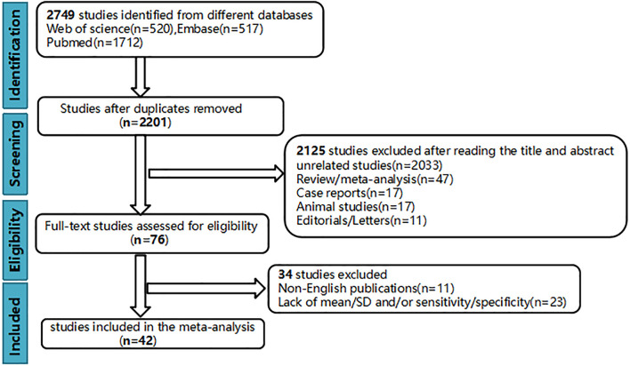
PubMed, Web of Science, and Embase databases were searched for relevant studies published from inception to October 2021. Review Manager 5.3 was used to summarize standardized mean differences (SMDs) of mean kurtosis (MK), mean diffusivity (MD), tissue diffusivity (D), pseudo diffusivity (D*), perfusion fraction (f), mean apparent diffusion coefficient (ADCmean), and minimum apparent diffusion coefficient (ADCmin). Stata12.0 was used to pool the sensitivity, specificity, and area under the curve (AUC). Overall, 42 up-to-standard studies with 3,807 cases of HCC were included in the meta-analysis.

RESULTS

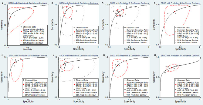
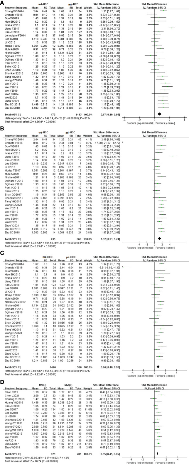
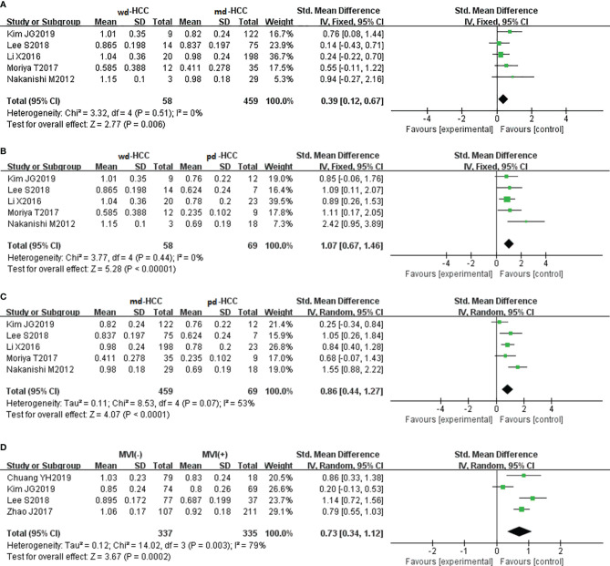
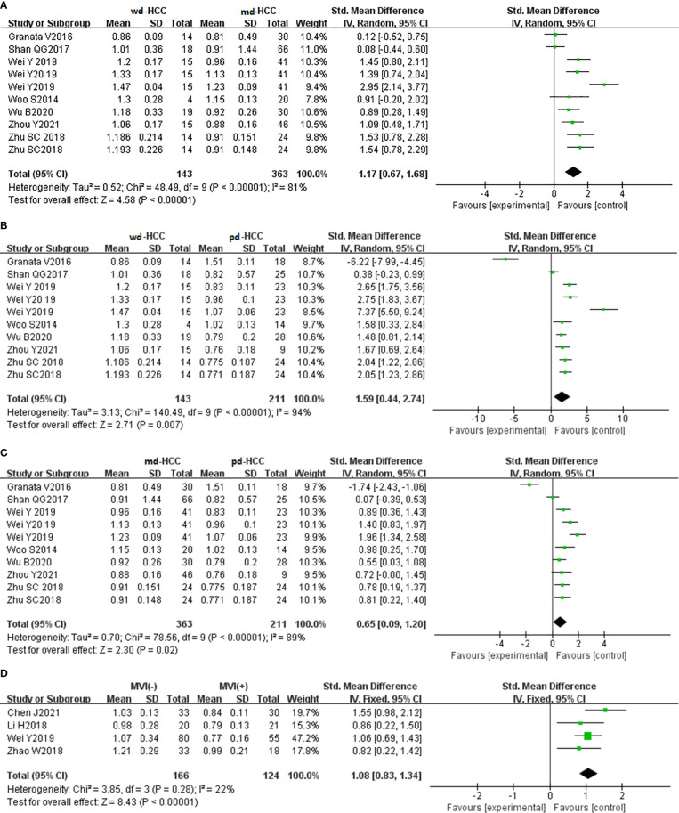
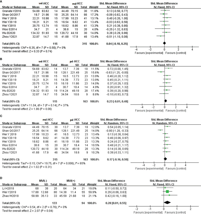
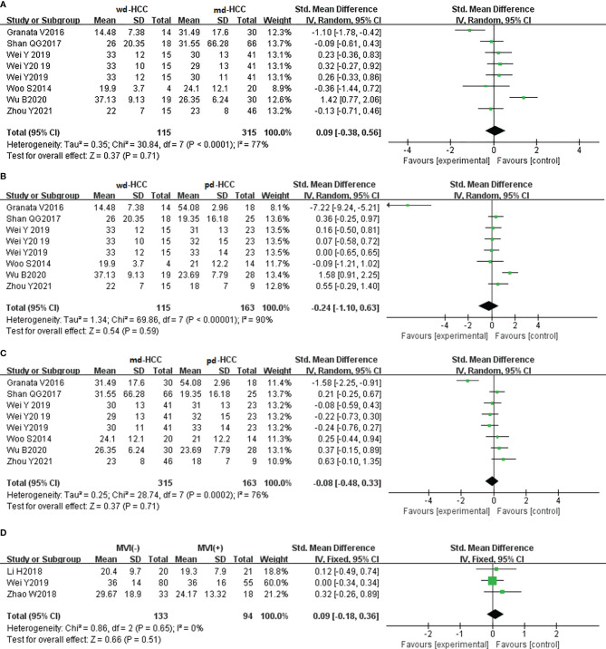
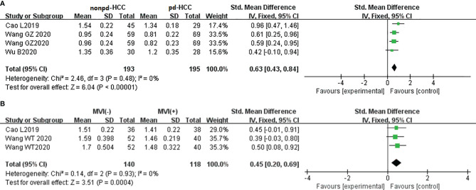
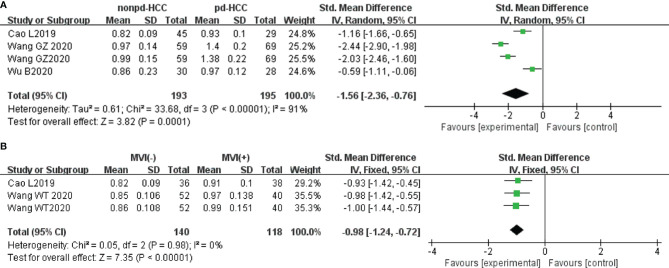
The SMDs of ADCmean, ADCmin, and D values, but not those of D* and f values, significantly differed between well, moderately, and poorly differentiated HCC ( < 0.01). The sensitivity, specificity, and AUC of the MK, D, ADCmean, and ADCmin for preoperative prediction of poorly differentiated HCC were 69%/94%/0.89, 87%/80%/0.89, 82%/75%/0.86, and 83%/64%/0.81, respectively. In addition, the sensitivity, specificity, and AUC of the D and ADCmean for preoperative prediction of well-differentiated HCC were 87%/83%/0.92 and 82%/88%/0.90, respectively. The SMDs of ADCmean, ADCmin, D, MD, and MK values, but not f values, showed significant differences ( < 0.01) between MVI-positive (MVI+) and MVI-negative (MVI-) HCC. The sensitivity and specificity of D and ADCmean for preoperative prediction of MVI+ were 80%/80% and 74%/71%, respectively; the AUC of the D (0.87) was significantly higher than that of ADCmean (0.78) ( = -2.208, = 0.027). Sensitivity analysis showed that the results of the above parameters were stable and reliable, and subgroup analysis confirmed a good prediction effect.

CONCLUSION

DKI parameters (MD and MK) and IVIM-DWI parameters (D value, ADCmean, and ADCmin) can be used as a noninvasive and simple preoperative examination method to predict the grade and MVI in HCC. Compared with ADCmean and ADCmin, MD and D values have higher diagnostic efficacy in predicting the grades of HCC, and D value has superior diagnostic efficacy to ADCmean in predicting MVI+ in HCC. However, f value cannot predict the grade or MVI in HCC.

摘要

背景

目前,在预测肝细胞癌(HCC)的病理分级和微血管侵犯(MVI)方面,扩散峰度成像(DKI)、体素内不相干运动(IVIM)和扩散加权成像(DWI)的参数存在争议。本研究的目的是调查和比较DKI和IVIM-DWI参数对HCC病理分级和MVI术前评估的预测能力。

方法

检索PubMed、Web of Science和Embase数据库中从建库至2021年10月发表的相关研究。使用Review Manager 5.3总结平均峰度(MK)、平均扩散率(MD)、组织扩散率(D)、伪扩散率(D*)、灌注分数(f)、平均表观扩散系数(ADCmean)和最小表观扩散系数(ADCmin)的标准化均数差(SMD)。使用Stata12.0汇总敏感性、特异性和曲线下面积(AUC)。总体而言,42项符合标准的研究共3807例HCC患者被纳入荟萃分析。

结果

在高分化、中分化和低分化HCC之间,ADCmean、ADCmin和D值的SMD有显著差异(<0.01),而D*和f值的SMD无显著差异。MK、D、ADCmean和ADCmin术前预测低分化HCC的敏感性、特异性和AUC分别为69%/94%/0.89、87%/80%/0.89、82%/75%/0.86和83%/64%/0.81。此外,D和ADCmean术前预测高分化HCC的敏感性、特异性和AUC分别为87%/83%/0.92和82%/88%/0.90。在MVI阳性(MVI+)和MVI阴性(MVI-)的HCC之间,ADCmean、ADCmin、D、MD和MK值的SMD有显著差异(<0.01),而f值的SMD无显著差异。D和ADCmean术前预测MVI+的敏感性和特异性分别为80%/80%和74%/71%;D的AUC(0.87)显著高于ADCmean的AUC(0.78)(z=-2.208,P=0.027)。敏感性分析表明上述参数结果稳定可靠,亚组分析证实预测效果良好。

结论

DKI参数(MD和MK)和IVIM-DWI参数(D值、ADCmean和ADCmin)可作为一种无创且简便的术前检查方法来预测HCC的分级和MVI。与ADCmean和ADCmin相比,MD和D值在预测HCC分级方面具有更高的诊断效能,D值在预测HCC的MVI+方面比ADCmean具有更高的诊断效能。然而,f值无法预测HCC的分级或MVI。

https://cdn.ncbi.nlm.nih.gov/pmc/blobs/d8c7/9131658/794dcfa015c4/fonc-12-884854-g010.jpg
https://cdn.ncbi.nlm.nih.gov/pmc/blobs/d8c7/9131658/93745033f5dc/fonc-12-884854-g001.jpg
https://cdn.ncbi.nlm.nih.gov/pmc/blobs/d8c7/9131658/a0288d1a7b92/fonc-12-884854-g002.jpg
https://cdn.ncbi.nlm.nih.gov/pmc/blobs/d8c7/9131658/d1d84a85102f/fonc-12-884854-g003.jpg
https://cdn.ncbi.nlm.nih.gov/pmc/blobs/d8c7/9131658/87b8fd5ec00d/fonc-12-884854-g004.jpg
https://cdn.ncbi.nlm.nih.gov/pmc/blobs/d8c7/9131658/c6681c5e80d9/fonc-12-884854-g005.jpg
https://cdn.ncbi.nlm.nih.gov/pmc/blobs/d8c7/9131658/2818709b43d7/fonc-12-884854-g006.jpg
https://cdn.ncbi.nlm.nih.gov/pmc/blobs/d8c7/9131658/1847a8834ba2/fonc-12-884854-g007.jpg
https://cdn.ncbi.nlm.nih.gov/pmc/blobs/d8c7/9131658/0984d79cf22b/fonc-12-884854-g008.jpg
https://cdn.ncbi.nlm.nih.gov/pmc/blobs/d8c7/9131658/7b4e5316f9d2/fonc-12-884854-g009.jpg
https://cdn.ncbi.nlm.nih.gov/pmc/blobs/d8c7/9131658/794dcfa015c4/fonc-12-884854-g010.jpg

相似文献

1
The Roles of Diffusion Kurtosis Imaging and Intravoxel Incoherent Motion Diffusion-Weighted Imaging Parameters in Preoperative Evaluation of Pathological Grades and Microvascular Invasion in Hepatocellular Carcinoma.扩散峰度成像和体素内不相干运动扩散加权成像参数在肝细胞癌病理分级和微血管侵犯术前评估中的作用
Front Oncol. 2022 May 11;12:884854. doi: 10.3389/fonc.2022.884854. eCollection 2022.
2
Quantitative analysis for detection and grading of hepatocellular carcinoma: Comparison of diffusion kurtosis imaging, intravoxel incoherent motion and conventional diffusion-weighted imaging.肝细胞癌检测与分级的定量分析:扩散峰度成像、体素内不相干运动与传统扩散加权成像的比较
Oncol Lett. 2022 Sep 22;24(5):403. doi: 10.3892/ol.2022.13523. eCollection 2022 Nov.
3
Diffusion kurtosis imaging (DKI) of hepatocellular carcinoma: correlation with microvascular invasion and histologic grade.肝细胞癌的扩散峰度成像(DKI):与微血管侵犯及组织学分级的相关性
Quant Imaging Med Surg. 2019 Apr;9(4):590-602. doi: 10.21037/qims.2019.02.14.
4
Diffusion-Weighted Imaging Reflects Tumor Grading and Microvascular Invasion in Hepatocellular Carcinoma.扩散加权成像反映肝细胞癌的肿瘤分级和微血管侵犯。
Liver Cancer. 2021 Feb;10(1):10-24. doi: 10.1159/000511384. Epub 2021 Jan 27.
5
Preoperative histogram analysis of intravoxel incoherent motion (IVIM) for predicting microvascular invasion in patients with single hepatocellular carcinoma.术前体素内不相干运动(IVIM)直方图分析预测单发肝细胞癌患者微血管侵犯。
Eur J Radiol. 2018 Aug;105:65-71. doi: 10.1016/j.ejrad.2018.05.032. Epub 2018 Jun 1.
6
Application of DKI and IVIM imaging in evaluating histologic grades and clinical stages of clear cell renal cell carcinoma.扩散峰度成像(DKI)和体素内不相干运动成像(IVIM)在评估透明细胞肾细胞癌组织学分级和临床分期中的应用。
Front Oncol. 2023 Oct 26;13:1203922. doi: 10.3389/fonc.2023.1203922. eCollection 2023.
7
Intravoxel incoherent motion and enhanced T2*-weighted angiography for preoperative prediction of microvascular invasion in hepatocellular carcinoma.体素内不相干运动及增强T2*加权血管造影术在肝细胞癌微血管侵犯术前预测中的应用
Front Oncol. 2024 Aug 9;14:1389769. doi: 10.3389/fonc.2024.1389769. eCollection 2024.
8
Volumetric analysis of intravoxel incoherent motion diffusion-weighted imaging in preoperative assessment of non-small cell lung cancer.基于体素内不相干运动扩散加权成像的术前评估非小细胞肺癌的容积分析。
Jpn J Radiol. 2022 Sep;40(9):903-913. doi: 10.1007/s11604-022-01279-w. Epub 2022 May 4.
9
Diffusion and perfusion MR parameters to assess preoperative short-course radiotherapy response in locally advanced rectal cancer: a comparative explorative study among Standardized Index of Shape by DCE-MRI, intravoxel incoherent motion- and diffusion kurtosis imaging-derived parameters.扩散和灌注磁共振参数评估局部进展期直肠癌术前短程放疗反应:DCE-MRI 标准化形状指数、体素内不相干运动和扩散峰度成像衍生参数的比较探索性研究。
Abdom Radiol (NY). 2019 Nov;44(11):3683-3700. doi: 10.1007/s00261-018-1801-z.
10
The Benefit of Apparent Diffusion Coefficient in Evaluating the Invasiveness of Hepatocellular Carcinoma.表观扩散系数在评估肝细胞癌侵袭性中的作用
Front Oncol. 2021 Aug 24;11:719480. doi: 10.3389/fonc.2021.719480. eCollection 2021.

引用本文的文献

1
Chinese expert consensus on the diagnosis and treatment of hepatocellular carcinoma with microvascular invasion (2024 edition).肝细胞癌微血管侵犯诊断与治疗中国专家共识(2024年版)
Hepatobiliary Surg Nutr. 2025 Apr 1;14(2):246-266. doi: 10.21037/hbsn-24-359. Epub 2024 Dec 30.
2
Clinicopathologic and ultrasonographic features of combined hepatocellular-cholangiocarcinoma and its correlation with microvascular invasion: a predictive role of contrast-enhanced ultrasound.肝细胞-胆管细胞癌的临床病理及超声特征及其与微血管侵犯的相关性:超声造影的预测作用
Front Oncol. 2024 Dec 20;14:1474675. doi: 10.3389/fonc.2024.1474675. eCollection 2024.
3

本文引用的文献

1
Preoperative assessment of microvascular invasion of hepatocellular carcinoma using non-Gaussian diffusion-weighted imaging with a fractional order calculus model: A pilot study.应用分数阶微积分模型的非高斯扩散加权成像对肝细胞癌微血管侵犯的术前评估:一项初步研究。
Magn Reson Imaging. 2023 Jan;95:110-117. doi: 10.1016/j.mri.2021.09.003. Epub 2021 Sep 8.
2
The Role of Non-Gaussian Models of Diffusion Weighted MRI in Hepatocellular Carcinoma: A Systematic Review.扩散加权磁共振成像的非高斯模型在肝细胞癌中的作用:一项系统评价
J Clin Med. 2021 Jun 15;10(12):2641. doi: 10.3390/jcm10122641.
3
A study of the correlations between IVIM-DWI parameters and the histologic differentiation of hepatocellular carcinoma.
Nomogram based on the neutrophil-to-lymphocyte ratio and MR diffusion quantitative parameters for predicting Ki67 expression in hepatocellular carcinoma from a prospective study.
基于中性粒细胞与淋巴细胞比值和磁共振扩散定量参数的列线图,用于预测一项前瞻性研究中肝细胞癌的Ki67表达。
Sci Rep. 2024 Dec 30;14(1):31738. doi: 10.1038/s41598-024-82333-7.
4
Preoperative prediction of microvascular invasion: new insights into personalized therapy for early-stage hepatocellular carcinoma.微血管侵犯的术前预测:早期肝细胞癌个体化治疗的新见解
Quant Imaging Med Surg. 2024 Jul 1;14(7):5205-5223. doi: 10.21037/qims-24-44. Epub 2024 Jun 27.
5
Dual-energy computed tomography iodine quantification combined with laboratory data for predicting microvascular invasion in hepatocellular carcinoma: a two-centre study.双能 CT 碘定量与实验室数据联合预测肝细胞癌微血管侵犯的价值:一项多中心研究。
Br J Radiol. 2024 Aug 1;97(1160):1467-1475. doi: 10.1093/bjr/tqae116.
6
The value of varying diffusion curvature MRI for assessing the microvascular invasion of hepatocellular carcinoma.弥散曲率 MRI 对评估肝细胞癌微血管侵犯的价值。
Abdom Radiol (NY). 2024 Apr;49(4):1154-1164. doi: 10.1007/s00261-023-04168-6. Epub 2024 Feb 5.
7
The Feasibility of Using Tri-Exponential Intra-Voxel Incoherent Motion DWI for Identifying the Microvascular Invasion in Hepatocellular Carcinoma.使用三指数体素内不相干运动扩散加权成像识别肝细胞癌微血管侵犯的可行性
J Hepatocell Carcinoma. 2023 Sep 29;10:1659-1671. doi: 10.2147/JHC.S433948. eCollection 2023.
8
Radiological findings in non-surgical recurrent hepatocellular carcinoma: From locoregional treatments to immunotherapy.非手术复发性肝细胞癌的放射学表现:从局部区域治疗到免疫治疗。
World J Gastroenterol. 2023 Mar 21;29(11):1669-1684. doi: 10.3748/wjg.v29.i11.1669.
9
Differential value of diffusion kurtosis imaging and intravoxel incoherent motion in benign and malignant solitary pulmonary lesions.扩散峰度成像和体素内不相干运动在良恶性孤立性肺病变中的鉴别价值。
Front Oncol. 2023 Jan 12;12:1075072. doi: 10.3389/fonc.2022.1075072. eCollection 2022.
10
Clinical and imaging features preoperative evaluation of histological grade and microvascular infiltration of hepatocellular carcinoma.术前评估肝细胞癌的组织学分级和微血管浸润的临床和影像学特征。
BMC Gastroenterol. 2022 Aug 1;22(1):369. doi: 10.1186/s12876-022-02449-w.
一项研究探讨了 IVIM-DWI 参数与肝细胞癌组织学分化之间的相关性。
Sci Rep. 2021 May 17;11(1):10392. doi: 10.1038/s41598-021-89784-2.
4
Diffusion-Weighted Imaging Reflects Tumor Grading and Microvascular Invasion in Hepatocellular Carcinoma.扩散加权成像反映肝细胞癌的肿瘤分级和微血管侵犯。
Liver Cancer. 2021 Feb;10(1):10-24. doi: 10.1159/000511384. Epub 2021 Jan 27.
5
Global Cancer Statistics 2020: GLOBOCAN Estimates of Incidence and Mortality Worldwide for 36 Cancers in 185 Countries.《全球癌症统计数据 2020:全球 185 个国家和地区 36 种癌症的发病率和死亡率估计》。
CA Cancer J Clin. 2021 May;71(3):209-249. doi: 10.3322/caac.21660. Epub 2021 Feb 4.
6
Prediction of Microvascular Invasion of Hepatocellular Carcinoma Based on Preoperative Diffusion-Weighted MR Using Deep Learning.基于深度学习的术前磁共振弥散加权成像预测肝细胞癌微血管侵犯。
Acad Radiol. 2021 Nov;28 Suppl 1:S118-S127. doi: 10.1016/j.acra.2020.11.014. Epub 2020 Dec 7.
7
Amide Proton Transfer Imaging vs Diffusion Kurtosis Imaging for Predicting Histological Grade of Hepatocellular Carcinoma.酰胺质子转移成像与扩散峰度成像对预测肝细胞癌组织学分级的比较
J Hepatocell Carcinoma. 2020 Oct 9;7:159-168. doi: 10.2147/JHC.S272535. eCollection 2020.
8
Evaluation of Multiple Prognostic Factors of Hepatocellular Carcinoma with Intra-Voxel Incoherent Motions Imaging by Extracting the Histogram Metrics.通过提取直方图指标评估体素内不相干运动成像对肝细胞癌多个预后因素的影响。
Cancer Manag Res. 2020 Jul 20;12:6019-6031. doi: 10.2147/CMAR.S262973. eCollection 2020.
9
Magnetic Resonance Diffusion Kurtosis Imaging versus Diffusion-Weighted Imaging in Evaluating the Pathological Grade of Hepatocellular Carcinoma.磁共振扩散峰度成像与扩散加权成像在评估肝细胞癌病理分级中的应用
Cancer Manag Res. 2020 Jun 29;12:5147-5158. doi: 10.2147/CMAR.S254371. eCollection 2020.
10
Diffusion-weighted imaging for identifying patients at high risk of tumor recurrence following liver transplantation.弥散加权成像识别肝移植后肿瘤复发高危患者。
Cancer Imaging. 2019 Nov 15;19(1):74. doi: 10.1186/s40644-019-0264-y.