Tan Zhi-Gang, Zhou Qian, Cui Yan, Yi Lei, Ouyang Yian, Jiang Yugang
Department of Neurosurgery, The Second Xiangya Hospital of Central South University (CSU), Changsha, Hunan, China.
Medicine (Baltimore). 2016 Jun;95(26):e4047. doi: 10.1097/MD.0000000000004047.
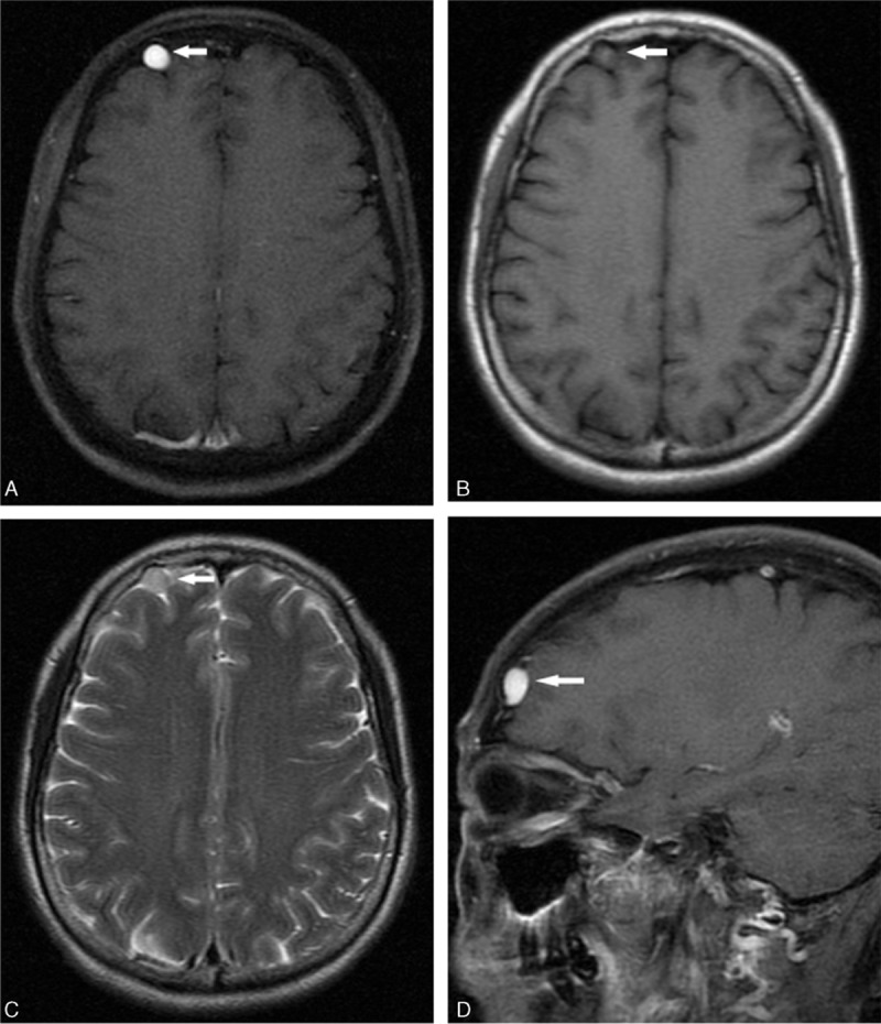
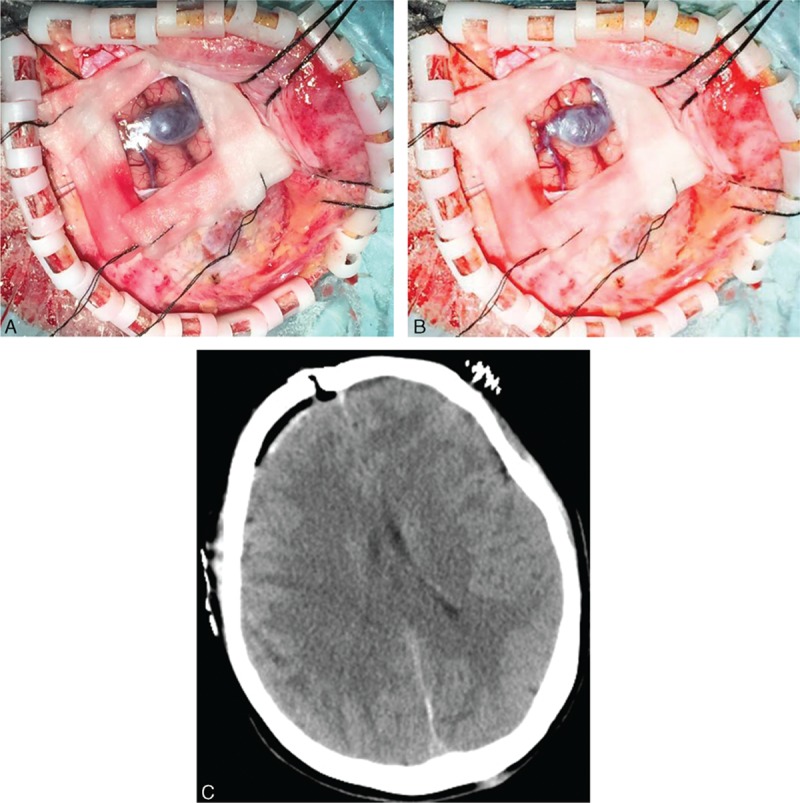
Isolated cerebral varix is a rare cerebrovascular anomaly, which is easily misdiagnosed as other brain tumors.A 59-year-old female patient with noncontributory medical history presented with headache and insomnia for the last 2 months. Upon admission, her neurological examination was unremarkable. Magnetic resonance imaging revealed a well-demarcated extra medullary mass, 11 × 11 mm in size, within the subdural space at the right frontal lobe. The lesion was initially interpreted as a convexity meningioma. After conducting a craniotomy on the patient, an extra-axial varix was exposed and resected subsequently. The patient's headache was resolved soon after surgery and charged without neurologic sequelae.Extra-axial isolated cerebral varix is mimicking convexity meningioma on MR images and should be considered as a differential diagnosis. The focal erosion in the inner table of the skull could be an important character of extra-axial isolated cerebral varix. An extremely round shape and smooth contour of the lesion was another important character. Isolated cerebral varix is rare vascular lesion that is treated surgically in the case of rupture or compression of adjacent structures. The information obtained with noninvasive imaging techniques should include CTA to make a clinical decision.
孤立性脑静脉曲张是一种罕见的脑血管畸形,很容易被误诊为其他脑肿瘤。一名59岁女性患者,既往病史无特殊,近2个月出现头痛和失眠。入院时,她的神经系统检查无异常。磁共振成像显示在右额叶硬膜下间隙有一个边界清晰的髓外肿块,大小为11×11毫米。该病变最初被诊断为凸面脑膜瘤。对患者进行开颅手术后,暴露并切除了一个轴外静脉曲张。患者术后头痛很快缓解,且未遗留神经后遗症。轴外孤立性脑静脉曲张在磁共振图像上类似凸面脑膜瘤,应作为鉴别诊断考虑。颅骨内板的局灶性侵蚀可能是轴外孤立性脑静脉曲张的一个重要特征。病变极其圆形的形状和光滑的轮廓是另一个重要特征。孤立性脑静脉曲张是一种罕见的血管病变,在破裂或压迫相邻结构时需手术治疗。通过非侵入性成像技术获得的信息应包括CTA以做出临床决策。