• 文献检索
  • 文档翻译
  • 深度研究
  • 学术资讯
  • Suppr Zotero 插件Zotero 插件
  • 邀请有礼
  • 套餐&价格
  • 历史记录
应用&插件
Suppr Zotero 插件Zotero 插件浏览器插件Mac 客户端Windows 客户端微信小程序
定价
高级版会员购买积分包购买API积分包
服务
文献检索文档翻译深度研究API 文档MCP 服务
关于我们
关于 Suppr公司介绍联系我们用户协议隐私条款
关注我们

Suppr 超能文献

核心技术专利:CN118964589B侵权必究
粤ICP备2023148730 号-1Suppr @ 2026

文献检索

告别复杂PubMed语法,用中文像聊天一样搜索,搜遍4000万医学文献。AI智能推荐,让科研检索更轻松。

立即免费搜索

文件翻译

保留排版,准确专业,支持PDF/Word/PPT等文件格式,支持 12+语言互译。

免费翻译文档

深度研究

AI帮你快速写综述,25分钟生成高质量综述,智能提取关键信息,辅助科研写作。

立即免费体验

EOS成像与传统放射成像:一项卫生技术评估研究。

EOS imaging versus current radiography: A health technology assessment study.

作者信息

Mahboub-Ahari Alireza, Hajebrahimi Sakineh, Yusefi Mahmoud, Velayati Ashraf

机构信息

PhD of Health Economics, Iranian Center of Excellence in Health Management, Department of Health Service Management, Tabriz University of Medical Sciences, Tabriz, Iran.

Professor of Urology, Iranian Center for Evidence-Based Medicine, Tabriz University of Medical Sciences, Tabriz, Iran.

出版信息

Med J Islam Repub Iran. 2016 Feb 17;30:331. eCollection 2016.

PMID:27390701
原文链接:https://pmc.ncbi.nlm.nih.gov/articles/PMC4898869/
Abstract

BACKGROUND

EOS is a 2D/3D muscle skeletal diagnostic imaging system. The device has been developed to produce a high quality 2D, full body radiographs in standing, sitting and squatting positions. Three dimensional images can be reconstructed via sterEOS software. This Health Technology Assessment study aimed to investigate efficacy, effectiveness and cost-effectiveness of new emerged EOS imaging system in comparison with conventional x-ray radiographic techniques.

METHODS

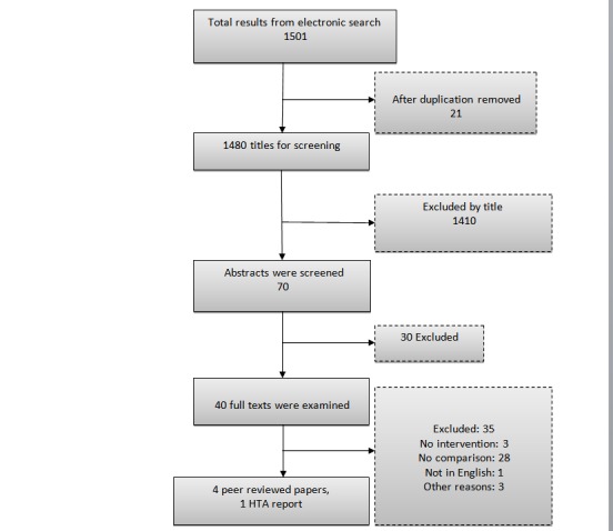
All cost and outcome data were assessed from Iran's Ministry of Health Perspective. Data for clinical effectiveness was extracted using a rigorous systematic review. As clinical outcomes the rate of x-ray emission and related quality of life were compared with Computed Radiography (CR) and Digital Radiography (DR). Standard costing method was conducted to find related direct medical costs. In order to examine robustness of the calculated Incremental Cost Effectiveness Ratios (ICERs) we used two-way sensitivity analysis. GDP Per capita of Islamic Republic of Iran (2012) adopted as cost-effectiveness threshold.

RESULTS

Review of related literature highlighted the lack of rigorous evidence for clinical outcomes. Ultra low dose EOS imaging device is known as a safe intervention because of FDA, CE and CSA certificates. The rate of emitted X-ray was 2 to 18 fold lower for EOS compared to the conventional techniques (p<0.001). The Incremental Cost Effectiveness Ratio for EOS relative to CR calculated $50706 in baseline analysis (the first scenario) and $50714, $9446 respectively for the second and third scenarios. Considering the value of neither $42146 as upper limit, nor the first neither the second scenario could pass the cost-effectiveness threshold for Iran.

CONCLUSION

EOS imaging technique might not be considered as a cost-effective intervention in routine practice of health system, especially within in-patient wards. Scenario analysis shows that, only in an optimum condition such as lower assembling costs and higher utilization rates, the device can be recruited for research and therapeutic purposes in pediatric orthopedic centers.

摘要

背景

EOS是一种二维/三维肌肉骨骼诊断成像系统。该设备旨在生成站立、坐姿和蹲姿下高质量的二维全身X光片。三维图像可通过sterEOS软件重建。这项卫生技术评估研究旨在调查新出现的EOS成像系统与传统X光射线照相技术相比的疗效、有效性和成本效益。

方法

所有成本和结果数据均从伊朗卫生部的角度进行评估。临床有效性数据通过严格的系统评价提取。作为临床结果,将X光发射率和相关生活质量与计算机X线摄影(CR)和数字X线摄影(DR)进行比较。采用标准成本核算方法来确定相关的直接医疗成本。为检验计算出的增量成本效益比(ICER)的稳健性,我们进行了双向敏感性分析。采用伊朗伊斯兰共和国2012年人均国内生产总值作为成本效益阈值。

结果

相关文献综述强调缺乏关于临床结果的严格证据。由于获得了美国食品药品监督管理局(FDA)、欧洲合格认证(CE)和加拿大标准协会(CSA)认证,超低剂量EOS成像设备被认为是一种安全的干预措施。与传统技术相比,EOS的X射线发射率低2至18倍(p<0.001)。在基线分析(第一种情况)中,EOS相对于CR的增量成本效益比计算为50706美元,在第二种和第三种情况中分别为50714美元和9446美元。考虑到42146美元既不是上限值,第一种和第二种情况都未达到伊朗的成本效益阈值。

结论

在卫生系统的常规实践中,尤其是在住院病房中,EOS成像技术可能不被视为具有成本效益的干预措施。情景分析表明,只有在诸如更低的组装成本和更高的利用率等最佳条件下,该设备才可在儿科骨科中心用于研究和治疗目的。

https://cdn.ncbi.nlm.nih.gov/pmc/blobs/81a9/4898869/963e1c429c83/mjiri-30-331-g001.jpg

相似文献

1
EOS imaging versus current radiography: A health technology assessment study.EOS成像与传统放射成像:一项卫生技术评估研究。
Med J Islam Repub Iran. 2016 Feb 17;30:331. eCollection 2016.
2
EOS 2D/3D X-ray imaging system: a systematic review and economic evaluation.EOS 2D/3D X 射线成像系统:系统评价和经济评估。
Health Technol Assess. 2012;16(14):1-188. doi: 10.3310/hta16140.
3
The EOS 2D/3D X-ray imaging system: a cost-effectiveness analysis quantifying the health benefits from reduced radiation exposure.EOS 2D/3D X 射线成像系统:一项成本效益分析,量化了降低辐射暴露带来的健康益处。
Eur J Radiol. 2013 Aug;82(8):e342-9. doi: 10.1016/j.ejrad.2013.02.015. Epub 2013 Mar 7.
4
Comparison of image quality using a X-ray stereotactical whole-body system and a direct flat-panel X-ray device in examinations of the pelvis and knee.使用X射线立体定向全身系统和直接平板X射线设备对骨盆和膝盖进行检查时的图像质量比较。
Rofo. 2014 Jan;186(1):67-76. doi: 10.1055/s-0033-1350441. Epub 2013 Sep 2.
5
Clinical validation of coronal and sagittal spinal curve measurements based on three-dimensional vertebra vector parameters.基于三维椎体矢量参数的冠状位和矢状位脊柱曲度测量的临床验证。
Spine J. 2012 Oct;12(10):960-8. doi: 10.1016/j.spinee.2012.08.175. Epub 2012 Sep 24.
6
EOS low-dose radiography: a reliable and accurate upright assessment of lower-limb lengths.EOS 低剂量射线摄影术:可靠且准确的下肢长度直立评估。
J Bone Joint Surg Am. 2013 Dec 4;95(23):e1831-7. doi: 10.2106/JBJS.L.00989.
7
Accuracy and reliability of coronal and sagittal spinal curvature data based on patient-specific three-dimensional models created by the EOS 2D/3D imaging system.基于 EOS 2D/3D 成像系统创建的患者特定三维模型的冠状和矢状脊柱曲率数据的准确性和可靠性。
Spine J. 2012 Nov;12(11):1052-9. doi: 10.1016/j.spinee.2012.10.002. Epub 2012 Oct 24.
8
Radiation dose of digital radiography (DR) versus micro-dose x-ray (EOS) on patients with adolescent idiopathic scoliosis: 2016 SOSORT- IRSSD "John Sevastic Award" Winner in Imaging Research.数字化X线摄影(DR)与微剂量X线(EOS)对青少年特发性脊柱侧凸患者的辐射剂量:2016年SOSORT-IRSSD影像学研究“约翰·塞瓦斯蒂克奖”获得者
Scoliosis Spinal Disord. 2016 Dec 29;11:46. doi: 10.1186/s13013-016-0106-7. eCollection 2016.
9
Comparison of 3-dimensional spinal reconstruction accuracy: biplanar radiographs with EOS versus computed tomography.三维脊柱重建精度的比较:EOS 与 CT 下双平面片。
Spine (Phila Pa 1976). 2012 Jul 15;37(16):1391-7. doi: 10.1097/BRS.0b013e3182518a15.
10
A systematic review of the clinical effectiveness of EOS 2D/3D X-ray imaging system.EOS 2D/3D X 射线成像系统临床效能的系统评价。
Eur Spine J. 2013 Feb;22(2):296-304. doi: 10.1007/s00586-012-2469-7. Epub 2012 Aug 19.

引用本文的文献

1
Health technology assessment in musculoskeletal radiology: the case study of EOSedge™.肌肉骨骼放射学中的卫生技术评估:EOSedgeTM 的案例研究。
Radiol Med. 2024 Jul;129(7):1076-1085. doi: 10.1007/s11547-024-01832-9. Epub 2024 Jun 10.
2
Pre-operative predictive factors of residual varus on the mechanical axis after Oxford unicompartmental knee arthroplasty.牛津单髁膝关节置换术后机械轴上残留内翻的术前预测因素。
Front Surg. 2023 Jan 9;9:1054351. doi: 10.3389/fsurg.2022.1054351. eCollection 2022.
3
The EOS 3D imaging system reliably measures posterior tibial slope.

本文引用的文献

1
Stated time preferences for health: a systematic review and meta analysis of private and social discount rates.健康的既定时间偏好:对个人和社会贴现率的系统评价与荟萃分析
J Res Health Sci. 2014 Summer;14(3):181-6.
2
EOS imaging of the human pelvis: reliability, validity, and controlled comparison with radiography.人体骨盆的 EOS 成像:可靠性、有效性和与射线照相术的对照研究。
J Bone Joint Surg Am. 2013 May 1;95(9):e58. doi: 10.2106/JBJS.K.01591.
3
Comparison of radiation dose, workflow, patient comfort and financial break-even of standard digital radiography and a novel biplanar low-dose X-ray system for upright full-length lower limb and whole spine radiography.
EOS 3D 成像系统能可靠地测量胫骨后倾角。
J Orthop Surg Res. 2021 Jun 16;16(1):388. doi: 10.1186/s13018-021-02529-9.
4
Accuracy of biplanar linear radiography versus conventional radiographs when used for lower limb and implant measurements.双平面直线放射摄影术与传统放射摄影术用于下肢和植入物测量时的准确性。
Arch Orthop Trauma Surg. 2022 May;142(5):735-745. doi: 10.1007/s00402-020-03700-3. Epub 2021 Jan 2.
5
Biplanar Low-Dose Radiography Is Accurate for Measuring Combined Anteversion After Total Hip Arthroplasty.双平面低剂量放射摄影术在测量全髋关节置换术后联合前倾角方面准确可靠。
HSS J. 2020 Feb;16(1):23-29. doi: 10.1007/s11420-018-09659-7. Epub 2019 Feb 5.
6
A Comparison of Patients Absorption Doses with Bone Deformity Due to the EOS Imaging and Digital Radiology.EOS成像与数字放射成像对伴有骨畸形患者吸收剂量的比较。
Arch Bone Jt Surg. 2017 May;5(3):145-148.
标准数字化放射摄影与新型双平面低剂量 X 射线系统在直立位全下肢和全脊柱摄影中的辐射剂量、工作流程、患者舒适度和经济收支平衡比较。
Skeletal Radiol. 2013 Jul;42(7):959-67. doi: 10.1007/s00256-013-1600-0. Epub 2013 Mar 28.
4
Comparison of scoliosis measurements based on three-dimensional vertebra vectors and conventional two-dimensional measurements: advantages in evaluation of prognosis and surgical results.基于三维椎体矢量与传统二维测量的脊柱侧弯测量比较:在评估预后和手术结果方面的优势。
Eur Spine J. 2013 Jun;22(6):1255-63. doi: 10.1007/s00586-012-2651-y. Epub 2013 Jan 23.
5
Musculoskeletal imaging in progress: the EOS imaging system.进行中的肌肉骨骼影像学:EOS 成像系统。
Joint Bone Spine. 2013 May;80(3):238-43. doi: 10.1016/j.jbspin.2012.09.018. Epub 2012 Nov 22.
6
Accuracy and reliability of coronal and sagittal spinal curvature data based on patient-specific three-dimensional models created by the EOS 2D/3D imaging system.基于 EOS 2D/3D 成像系统创建的患者特定三维模型的冠状和矢状脊柱曲率数据的准确性和可靠性。
Spine J. 2012 Nov;12(11):1052-9. doi: 10.1016/j.spinee.2012.10.002. Epub 2012 Oct 24.
7
New method for measuring acetabular component positioning with EOS imaging: feasibility study on dry bone.利用 EOS 成像测量髋臼部件定位的新方法:干骨的可行性研究。
Int Orthop. 2012 Nov;36(11):2205-9. doi: 10.1007/s00264-012-1650-2. Epub 2012 Sep 5.
8
Reliability of a new method for lower-extremity measurements based on stereoradiographic three-dimensional reconstruction.基于体层摄影三维重建的下肢测量新方法的可靠性。
Orthop Traumatol Surg Res. 2012 Sep;98(5):506-13. doi: 10.1016/j.otsr.2012.03.014. Epub 2012 Aug 1.
9
Recent advances in medical imaging: anatomical and clinical applications.医学成像的最新进展:解剖学与临床应用
Surg Radiol Anat. 2012 Oct;34(8):675-86. doi: 10.1007/s00276-012-0985-0. Epub 2012 May 29.
10
Posterior spinal fusion from T2 to the sacrum for the management of major deformities in patients with Parkinson disease: a retrospective review with analysis of complications.从T2至骶骨的后路脊柱融合术用于治疗帕金森病患者的严重畸形:一项并发症分析的回顾性研究
J Spinal Disord Tech. 2012 May;25(3):E53-60. doi: 10.1097/BSD.0b013e3182496670.