Hui Steve C N, Pialasse Jean-Philippe, Wong Judy Y H, Lam Tsz-Ping, Ng Bobby K W, Cheng Jack C Y, Chu Winnie C W
Department of Imaging and Interventional Radiology, Prince of Wales Hospital, The Chinese University of Hong Kong, Sha Tin, Hong Kong, SAR China.
Department of Imaging and Interventional Radiology, Prince of Wales Hospital, The Chinese University of Hong Kong, Sha Tin, Hong Kong, SAR China ; Department of Chiropractic, University of Quebec at Trois-Rivieres, Trois-Rivieres, Quebec Canada.
Scoliosis Spinal Disord. 2016 Dec 29;11:46. doi: 10.1186/s13013-016-0106-7. eCollection 2016.
Patients with adolescent idiopathic scoliosis (AIS) frequently receive x-ray imaging at diagnosis and subsequent follow monitoring. The ionizing radiation exposure has accumulated through their development stage and the effect of radiation to this young vulnerable group of patients is uncertain. To achieve the ALARA (as low as reasonably achievable) concept of radiation dose in medical imaging, a slot-scanning x-ray technique by the EOS system has been adopted and the radiation dose using micro-dose protocol was compared with the standard digital radiography on patients with AIS.
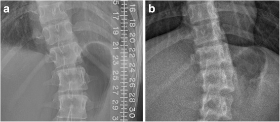
Ninety-nine participants with AIS underwent micro-dose EOS and 33 underwent standard digital radiography (DR) for imaging of the whole spine. Entrance-skin dose was measured using thermoluminescent dosimeters (TLD) at three regions (i.e. dorsal sites at the level of sternal notch, nipple line, symphysis pubis). Effective dose and organ dose were calculated by simulation using PCXMC 2.0. Data from two x-ray systems were compared using independent-samples t-test and significance level at 0.05. All TLD measurements were conducted on PA projection only. Image quality was also assessed by two raters using Cobb angle measurement and a set of imaging parameters for optimization purposes.
Entrance-skin dose from micro-dose EOS system was 5.9-27.0 times lower at various regions compared with standard DR. The calculated effective dose was 2.6 ± 0.5 (μSv) and 67.5 ± 23.3 (μSv) from micro-dose and standard DR, respectively. The reduction in the micro-dose was approximately 26 times. Organ doses at thyroid, lung and gonad regions were significantly lower in micro-dose ( < 0.001). Data were further compared within the different gender groups. Females received significantly higher ( < 0.001) organ dose at ovaries compared to the testes in males. Patients with AIS received approximately 16-34 times lesser organ dose from micro-dose x-ray as compared with the standard DR. There was no significant difference in overall rating of imaging quality between EOS and DR. Micro-dose protocol provided enough quality to perform consistent measurement on Cobb angle.
Entrance-skin dose, effective dose and organ dose were significantly reduced in micro-dose x-ray. The effective dose of a single micro-dose x-ray (2.6 μSv) was less than a day of background radiation. As AIS patients require periodic x-ray follow up for surveillance of curve progression, clinical use of micro-dose x-ray system is beneficial for these young patients to reduce the intake of ionizing radiation.
青少年特发性脊柱侧凸(AIS)患者在诊断及后续随访监测时经常接受X线成像检查。在其生长发育阶段,电离辐射暴露不断累积,而辐射对这群年轻易感患者的影响尚不确定。为在医学成像中实现辐射剂量的“合理尽可能低(ALARA)”理念,采用了EOS系统的狭缝扫描X线技术,并将微剂量方案的辐射剂量与AIS患者的标准数字X线摄影进行比较。
99名AIS参与者接受了微剂量EOS检查,33名接受了标准数字X线摄影(DR)以对整个脊柱进行成像。使用热释光剂量计(TLD)在三个区域(即胸骨切迹水平、乳头线、耻骨联合处的背部位置)测量体表入射剂量。使用PCXMC 2.0通过模拟计算有效剂量和器官剂量。使用独立样本t检验比较两个X线系统的数据,显著性水平为0.05。所有TLD测量仅在正位投照下进行。两名评估者还使用Cobb角测量和一组成像参数对图像质量进行评估以优化。
与标准DR相比,微剂量EOS系统在各个区域的体表入射剂量低5.9 - 27.0倍。微剂量和标准DR计算出的有效剂量分别为2.6±0.5(μSv)和67.5±23.3(μSv)。微剂量的降低幅度约为26倍。微剂量时甲状腺、肺和性腺区域的器官剂量显著更低(<0.001)。在不同性别组内进一步比较数据。女性卵巢的器官剂量显著高于男性睾丸(<0.001)。与标准DR相比,AIS患者接受微剂量X线检查时的器官剂量约低16 - 34倍。EOS和DR在成像质量的总体评分上无显著差异。微剂量方案提供了足够的质量以对Cobb角进行一致的测量。
微剂量X线显著降低了体表入射剂量、有效剂量和器官剂量。单次微剂量X线的有效剂量(2.6 μSv)低于一天的本底辐射。由于AIS患者需要定期进行X线随访以监测侧弯进展,微剂量X线系统的临床应用有利于这些年轻患者减少电离辐射的摄入。