Sandison Mairi E, Dempster John, McCarron John G
Strathclyde Institute of Pharmacy and Biomedical Sciences, University of Strathclyde, SIPBS Building, 161 Cathedral Street, Glasgow, G4 0RE, UK.
J Physiol. 2016 Nov 1;594(21):6189-6209. doi: 10.1113/JP272729. Epub 2016 Aug 13.
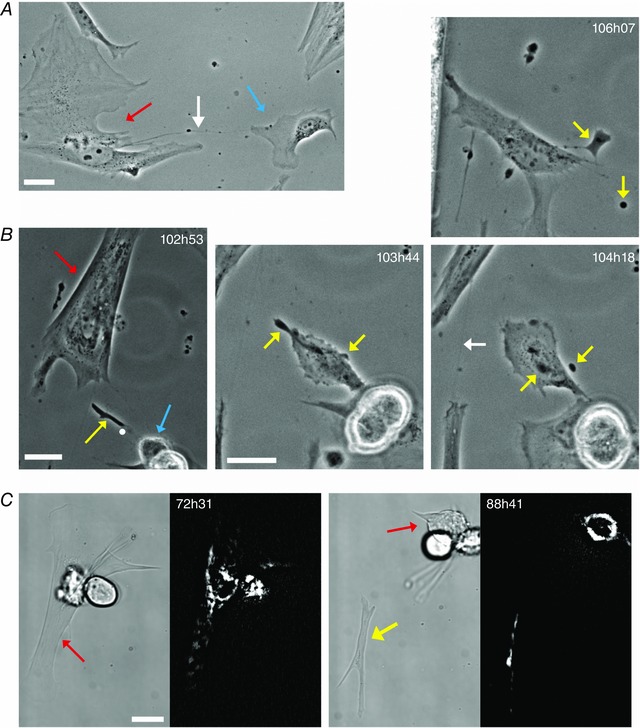
Smooth muscle cell (SMC) phenotypic conversion from a contractile to a migratory phenotype is proposed to underlie cardiovascular disease but its contribution to vascular remodelling and even its existence have recently been questioned. Tracking the fate of individual SMCs is difficult as no specific markers of migratory SMCs exist. This study used a novel, prolonged time-lapse imaging approach to continuously track the behaviour of unambiguously identified, fully differentiated SMCs. In response to serum, highly-elongated, contractile SMCs initially rounded up, before spreading and migrating and these migratory cells displayed clear phagocytic activity. This study provides a direct demonstration of the transition of fully contractile SMCs to a non-contractile, migratory phenotype with phagocytic capacity that may act as a macrophage-like cell.
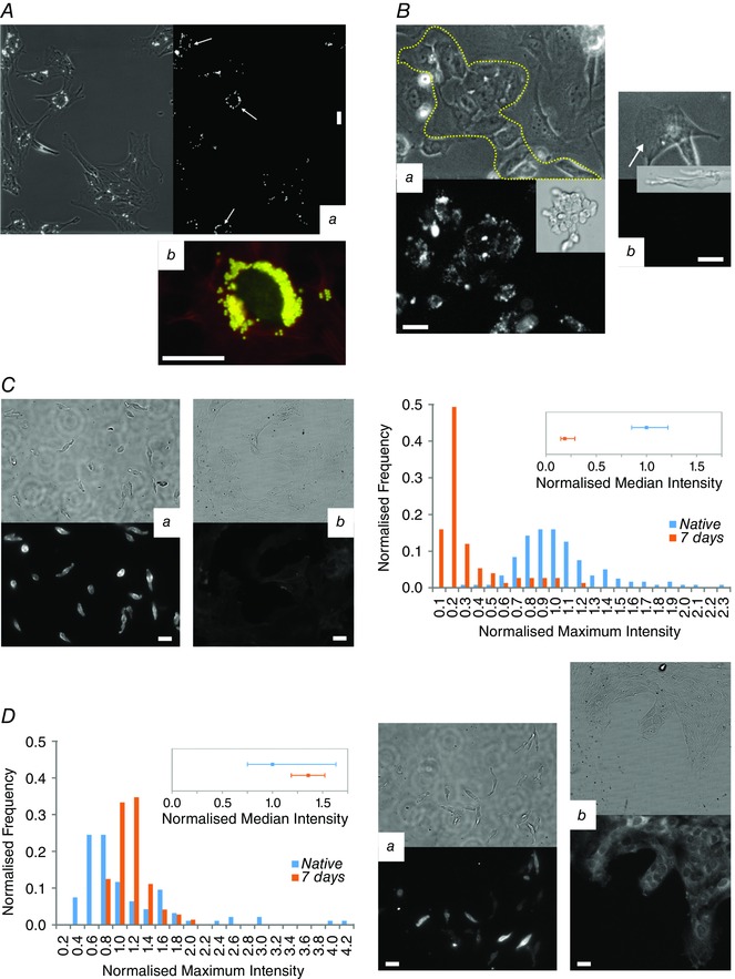
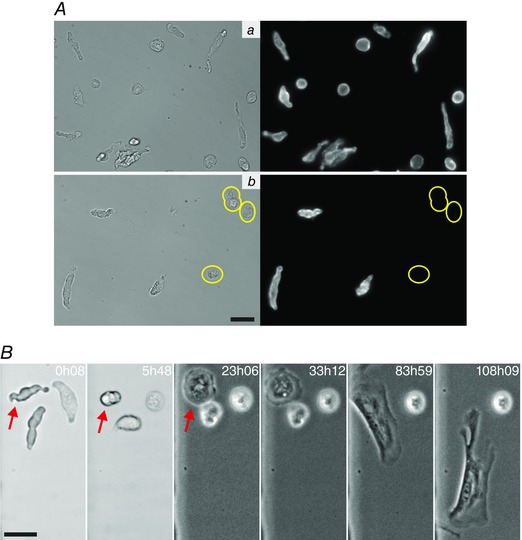
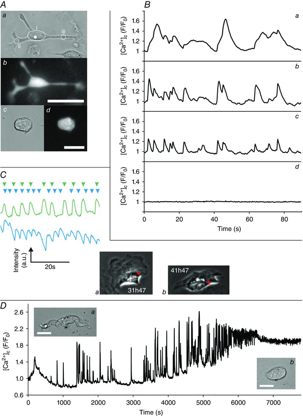
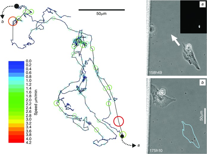
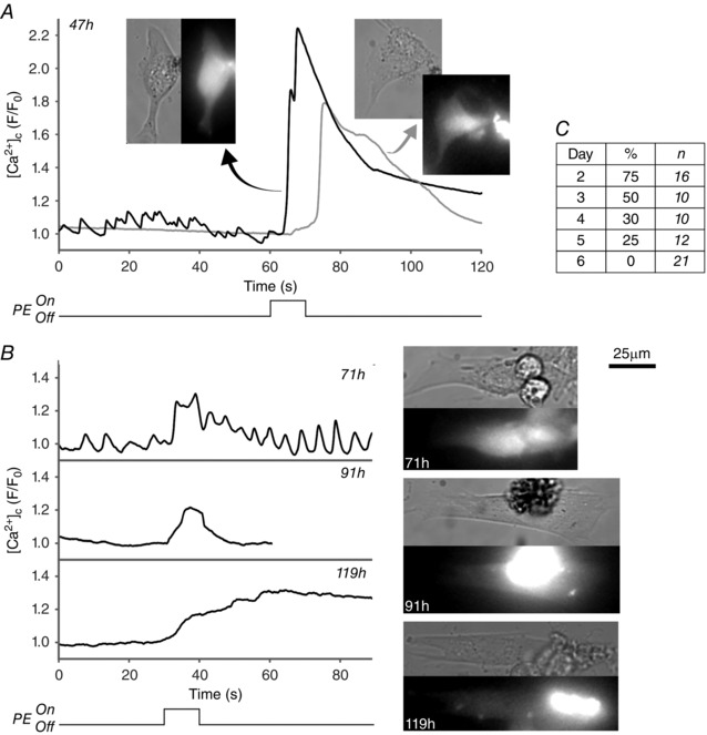
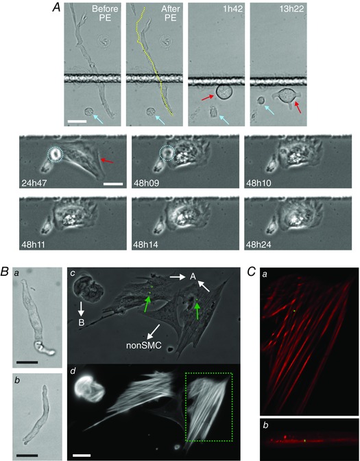
Atherosclerotic plaques are populated with smooth muscle cells (SMCs) and macrophages. SMCs are thought to accumulate in plaques because fully differentiated, contractile SMCs reprogramme into a 'synthetic' migratory phenotype, so-called phenotypic modulation, whilst plaque macrophages are thought to derive from blood-borne myeloid cells. Recently, these views have been challenged, with reports that SMC phenotypic modulation may not occur during vascular remodelling and that plaque macrophages may not be of haematopoietic origin. Following the fate of SMCs is complicated by the lack of specific markers for the migratory phenotype and direct demonstrations of phenotypic modulation are lacking. Therefore, we employed long-term, high-resolution, time-lapse microscopy to track the fate of unambiguously identified, fully-differentiated, contractile SMCs in response to the growth factors present in serum. Phenotypic modulation was clearly observed. The highly elongated, contractile SMCs initially rounded up, for 1-3 days, before spreading outwards. Once spread, the SMCs became motile and displayed dynamic cell-cell communication behaviours. Significantly, they also displayed clear evidence of phagocytic activity. This macrophage-like behaviour was confirmed by their internalisation of 1 μm fluorescent latex beads. However, migratory SMCs did not uptake acetylated low-density lipoprotein or express the classic macrophage marker CD68. These results directly demonstrate that SMCs may rapidly undergo phenotypic modulation and develop phagocytic capabilities. Resident SMCs may provide a potential source of macrophages in vascular remodelling.
平滑肌细胞(SMC)从收缩表型向迁移表型的转变被认为是心血管疾病的基础,但最近其对血管重塑的作用甚至其存在都受到了质疑。由于不存在迁移性SMC的特异性标志物,追踪单个SMC的命运很困难。本研究采用了一种新颖的、长时间延时成像方法来持续追踪明确鉴定的、完全分化的SMC的行为。响应血清时,高度伸长的收缩性SMC最初会变圆,然后伸展并迁移,这些迁移细胞表现出明显的吞噬活性。本研究直接证明了完全收缩性SMC向具有吞噬能力的非收缩性迁移表型的转变,这种表型可能起到类似巨噬细胞的作用。
动脉粥样硬化斑块中存在平滑肌细胞(SMC)和巨噬细胞。SMC被认为在斑块中积聚是因为完全分化的收缩性SMC重新编程为“合成”迁移表型,即所谓的表型调节,而斑块巨噬细胞被认为源自血源性髓样细胞。最近,这些观点受到了挑战,有报道称在血管重塑过程中SMC表型调节可能不会发生,并且斑块巨噬细胞可能不是造血起源。由于缺乏迁移表型的特异性标志物,追踪SMC的命运很复杂,并且缺乏表型调节的直接证据。因此,我们采用长期、高分辨率、延时显微镜来追踪明确鉴定的、完全分化的收缩性SMC在血清中生长因子作用下的命运。明显观察到了表型调节。高度伸长的收缩性SMC最初会变圆1 - 3天,然后向外伸展。一旦伸展,SMC变得具有运动性并表现出动态的细胞间通讯行为。重要的是,它们还表现出明显的吞噬活性证据。这种类似巨噬细胞的行为通过它们摄取1μm荧光乳胶珠得到证实。然而,迁移性SMC不摄取乙酰化低密度脂蛋白或表达经典巨噬细胞标志物CD68。这些结果直接证明了SMC可能迅速经历表型调节并发展出吞噬能力。驻留的SMC可能是血管重塑中巨噬细胞的一个潜在来源。