Division of Medical Genetics, Department of Internal Medicine, McGovern Medical School, The University of Texas Health Science Center at Houston, Houston, TX (A.C., C.S.K., K.K., A.K., J.C., D.M.M.).
Division of Cardiothoracic Surgery, Baylor College of Medicine, Houston, TX (.L., S.A.L., Y.H.S.).
Arterioscler Thromb Vasc Biol. 2021 Jan;41(1):302-316. doi: 10.1161/ATVBAHA.120.315164. Epub 2020 Oct 8.
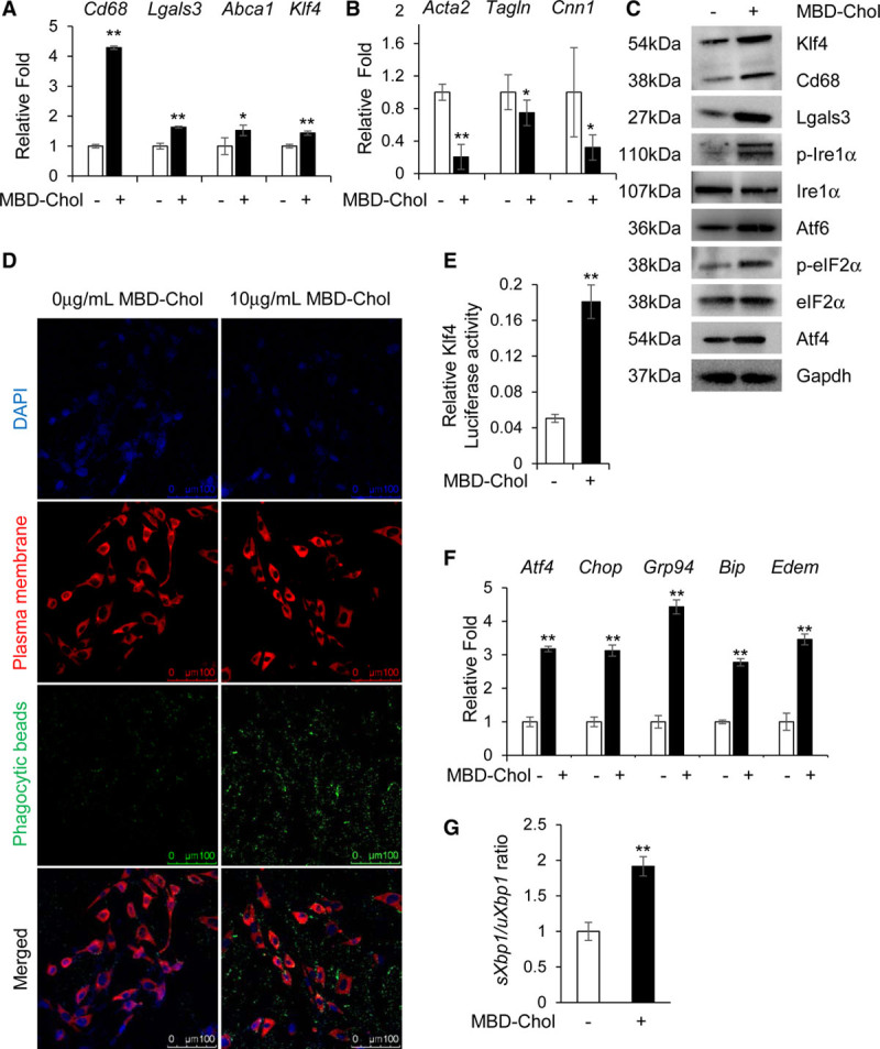
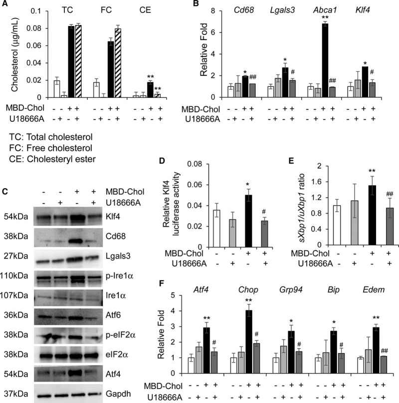
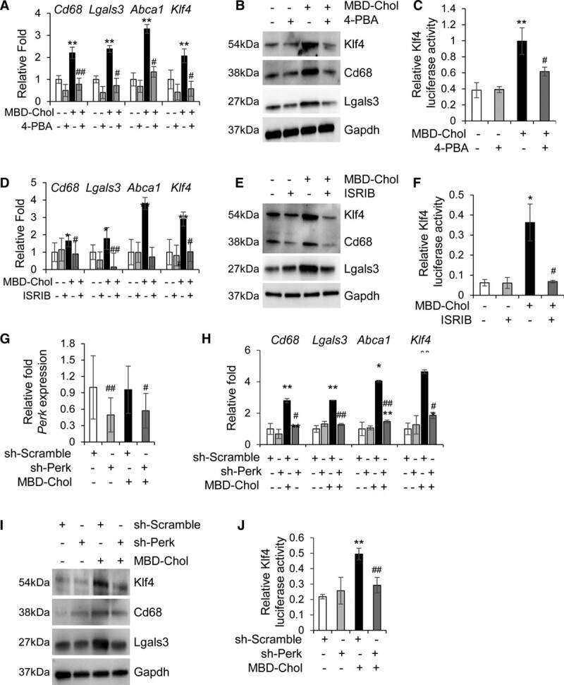
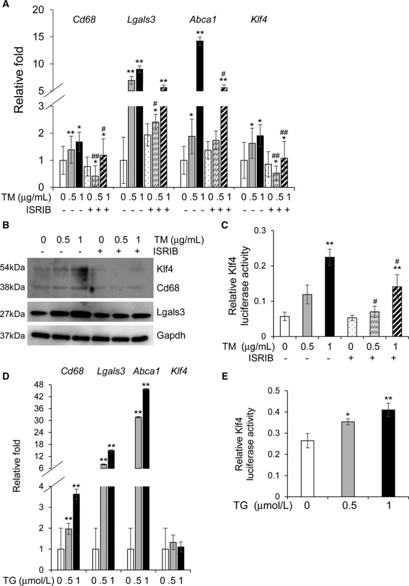
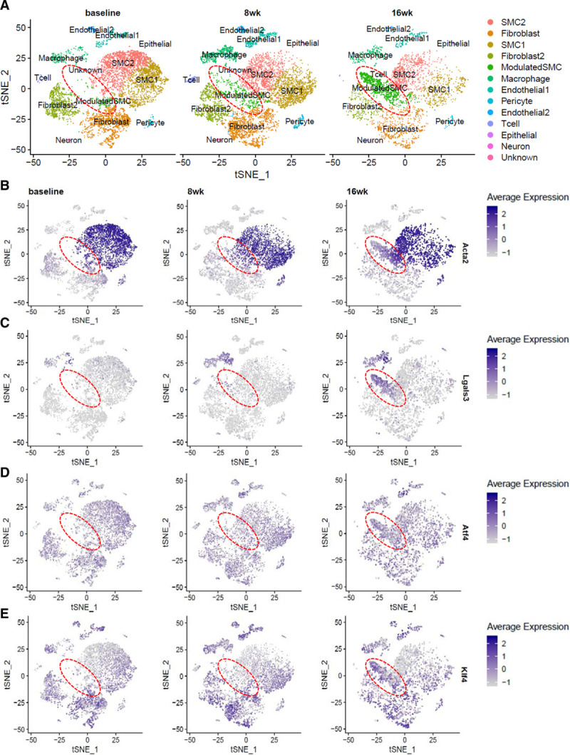
Vascular smooth muscle cells (SMCs) dedifferentiate and initiate expression of macrophage markers with cholesterol exposure. This phenotypic switching is dependent on the transcription factor Klf4 (Krüppel-like factor 4). We investigated the molecular pathway by which cholesterol induces SMC phenotypic switching. Approach and Results: With exposure to free cholesterol, SMCs decrease expression of contractile markers, activate Klf4, and upregulate a subset of macrophage and fibroblast markers characteristic of modulated SMCs that appear with atherosclerotic plaque formation. These phenotypic changes are associated with activation of all 3 pathways of the endoplasmic reticulum unfolded protein response (UPR), Perk (protein kinase RNA-like endoplasmic reticulum kinase), Ire (inositol-requiring enzyme) 1α, and Atf (activating transcription factor) 6. Blocking the movement of cholesterol from the plasma membrane to the endoplasmic reticulum prevents free cholesterol-induced UPR, Klf4 activation, and upregulation of the majority of macrophage and fibroblast markers. Cholesterol-induced phenotypic switching is also prevented by global UPR inhibition or specific inhibition of Perk signaling. Exposure to chemical UPR inducers, tunicamycin and thapsigargin, is sufficient to induce these same phenotypic transitions. Finally, analysis of published single-cell RNA sequencing data during atherosclerotic plaque formation in hyperlipidemic mice provides preliminary in vivo evidence of a role of UPR activation in modulated SMCs.
Our data demonstrate that UPR is necessary and sufficient to drive phenotypic switching of SMCs to cells that resemble modulated SMCs found in atherosclerotic plaques. Preventing a UPR in hyperlipidemic mice diminishes atherosclerotic burden, and our data suggest that preventing SMC transition to dedifferentiated cells expressing macrophage and fibroblast markers contributes to this decreased plaque burden.
血管平滑肌细胞(SMC)在胆固醇暴露下去分化并启动巨噬细胞标志物的表达。这种表型转换依赖于转录因子 Klf4(Krüppel 样因子 4)。我们研究了胆固醇诱导 SMC 表型转换的分子途径。
在暴露于游离胆固醇的情况下,SMC 会降低收缩标志物的表达,激活 Klf4,并上调一组与动脉粥样硬化斑块形成时出现的调制 SMC 特征相似的巨噬细胞和成纤维细胞标志物。这些表型变化与内质网未折叠蛋白反应(UPR)的所有 3 条途径的激活有关,包括 Perk(蛋白激酶 RNA 样内质网激酶)、Ire(肌醇需求酶)1α 和 Atf(激活转录因子)6。阻断胆固醇从质膜向内质网的运动可防止游离胆固醇诱导的 UPR、Klf4 激活以及大多数巨噬细胞和成纤维细胞标志物的上调。总体 UPR 抑制或 Perk 信号特异性抑制也可防止胆固醇诱导的表型转换。化学 UPR 诱导剂,如衣霉素和他普西隆,足以诱导这些相同的表型转变。最后,对高脂血症小鼠动脉粥样硬化斑块形成过程中发表的单细胞 RNA 测序数据的分析提供了 UPR 激活在调制 SMC 中的作用的初步体内证据。
我们的数据表明,UPR 是驱动 SMC 表型转换为类似于动脉粥样硬化斑块中发现的调制 SMC 的必需和充分条件。在高脂血症小鼠中阻止 UPR 可减少动脉粥样硬化负担,我们的数据表明,防止 SMC 向表达巨噬细胞和成纤维细胞标志物的去分化细胞转变有助于减少斑块负担。