Cautivo Kelly M, Lizama Carlos O, Tapia Pablo J, Agarwal Anil K, Garg Abhimanyu, Horton Jay D, Cortés Víctor A
Department of Nutrition, Diabetes and Metabolism, School of Medicine, Pontificia Universidad Católica de Chile, Santiago 8331150, Chile; Department of Molecular Genetics, University of Texas Southwestern Medical Center, Dallas, TX 75390, USA; Department of Internal Medicine, University of Texas Southwestern Medical Center, Dallas, TX 75390, USA.
Cardiovascular Research Institute, University of California, San Francisco, San Francisco, CA 94158, USA.
Mol Metab. 2016 May 13;5(7):491-505. doi: 10.1016/j.molmet.2016.05.004. eCollection 2016 Jul.
Characterize the cellular and molecular events responsible for lipodystrophy in AGPAT2 deficient mice.
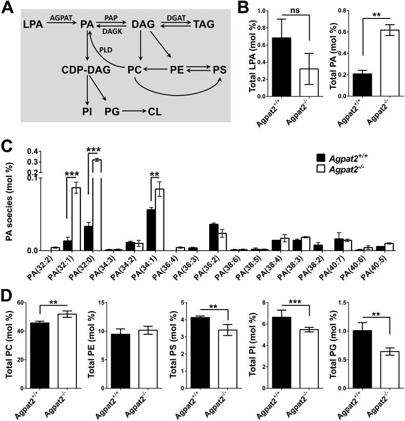
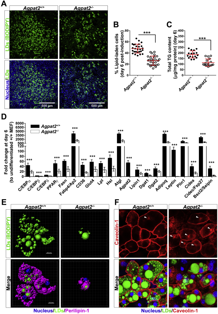
Adipose tissue and differentiated MEF were assessed using light and electron microscopy, followed by protein (immunoblots) and mRNA analysis (qPCR). Phospholipid profiling was determined by electrospray ionization tandem mass spectrometry (ESI-MS/MS).
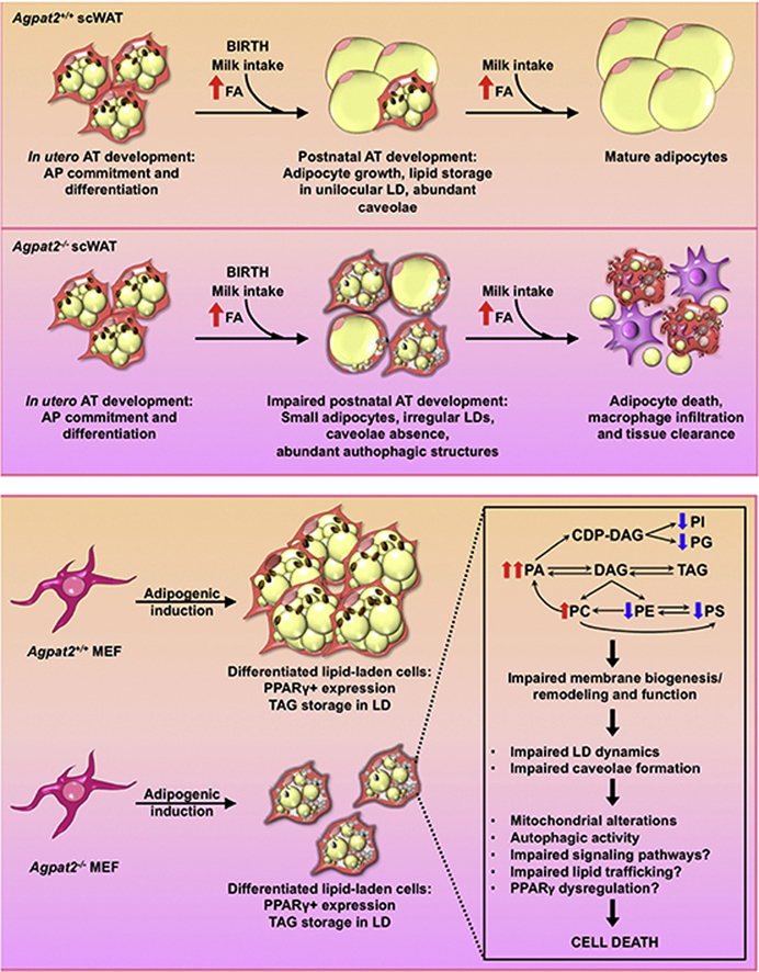
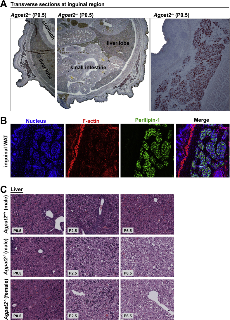
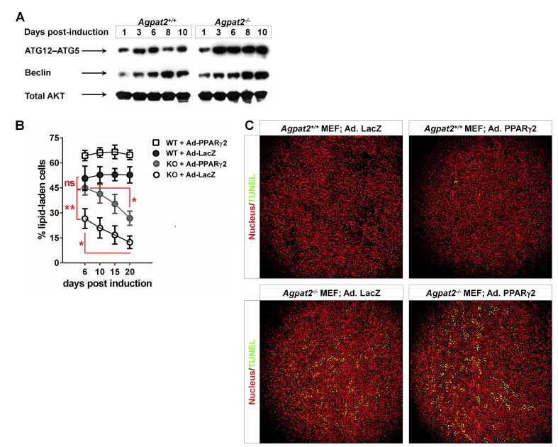
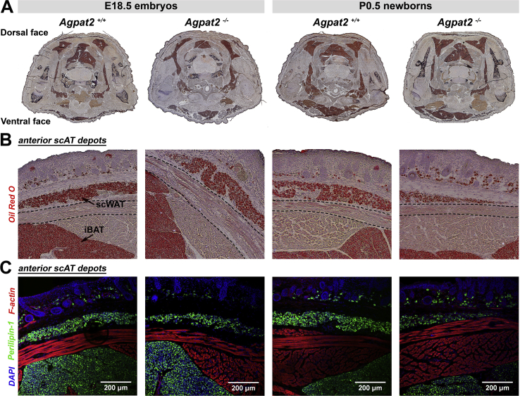
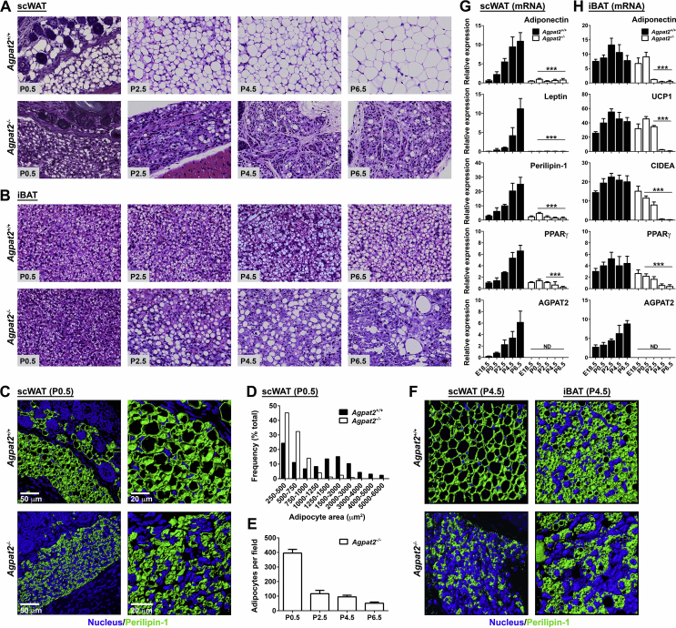
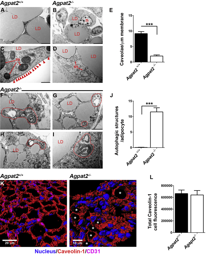
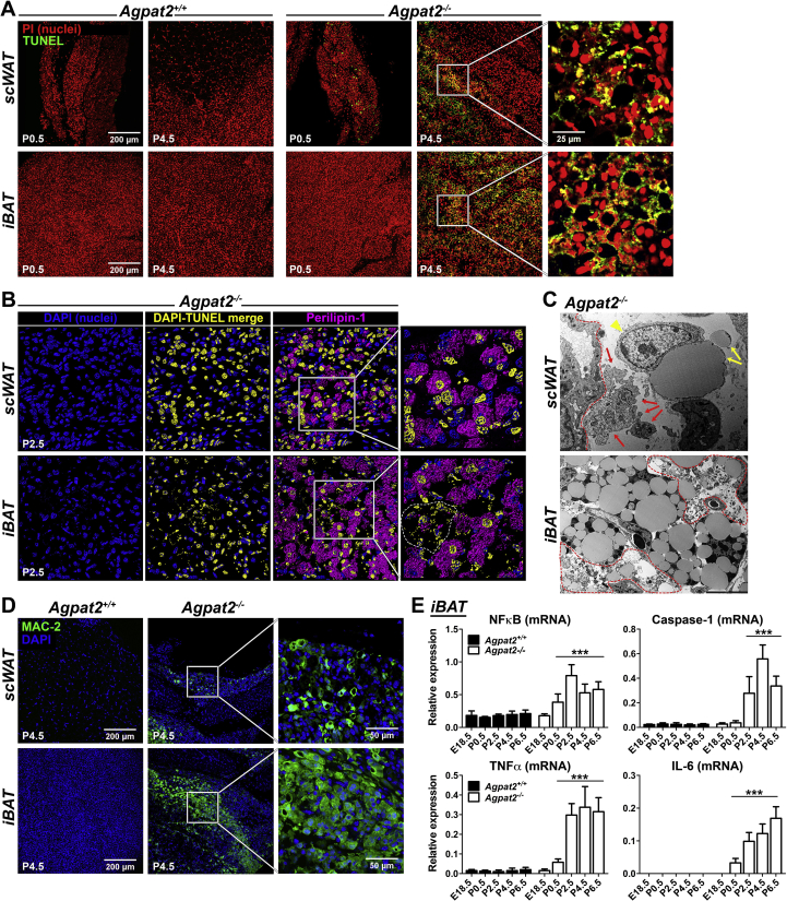
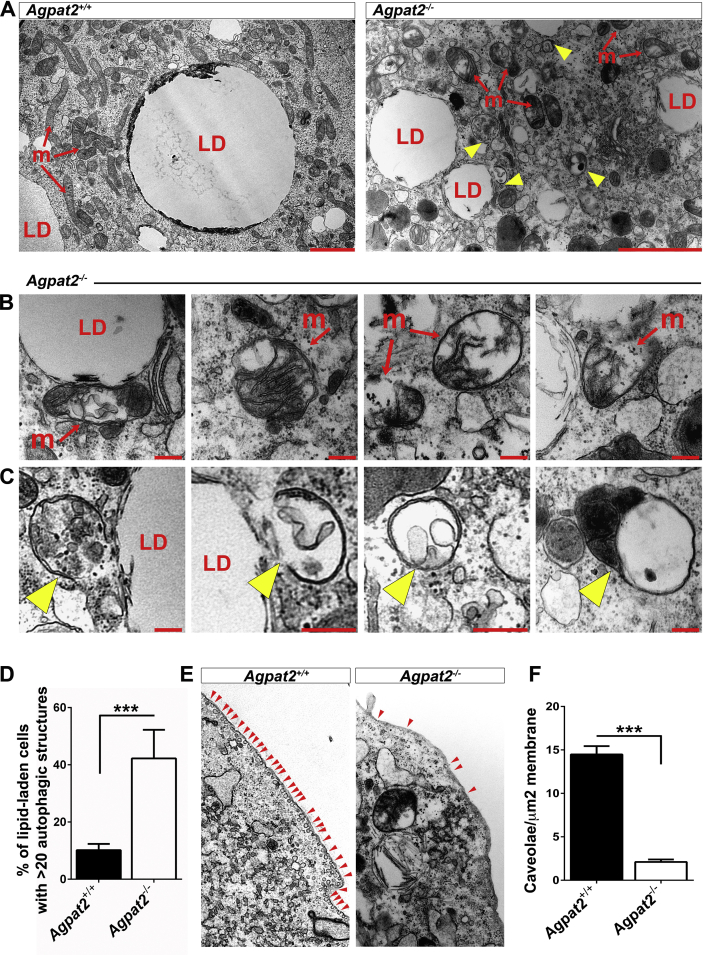
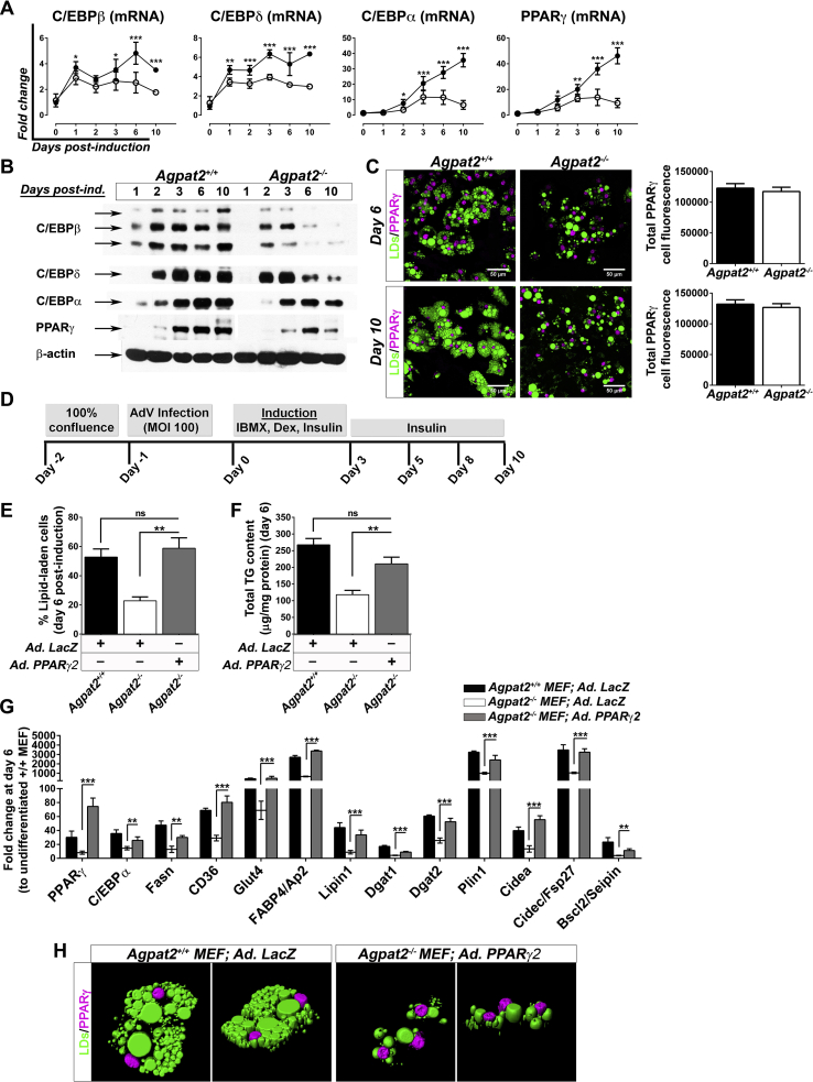
In contrast to adult Agpat2 (-/-) mice, fetuses and newborn Agpat2 (-/-) mice have normal mass of white and brown adipose tissue. Loss of both the adipose tissue depots occurs during the first week of postnatal life as a consequence of adipocyte death and inflammatory infiltration of the adipose tissue. At the ultrastructural level, adipose tissue of newborn Agpat2 (-/-) mice is virtually devoid of caveolae and has abnormal mitochondria and lipid droplets. Autophagic structures are also abundant. Consistent with these findings, differentiated Agpat2 (-/-) mouse embryonic fibroblasts (MEFs) also have impaired adipogenesis, characterized by a lower number of lipid-laden cells and ultrastructural abnormalities in lipid droplets, mitochondria and plasma membrane. Overexpression of PPARγ, the master regulator of adipogenesis, increased the number of Agpat2 (-/-) MEFs that differentiated into adipocyte-like cells but did not prevent morphological abnormalities and cell death. Furthermore, differentiated Agpat2 (-/-) MEFs have abnormal phospholipid compositions with 3-fold increased levels of phosphatidic acid.
We conclude that lipodystrophy in Agpat2 (-/-) mice results from postnatal cell death of adipose tissue in association with acute local inflammation. It is possible that AGPAT2 deficient adipocytes have an altered lipid filling or a reduced capacity to adapt the massive lipid availability associated with postnatal feeding.
确定AGPAT2基因缺陷小鼠脂肪营养不良相关的细胞和分子事件。
使用光学显微镜和电子显微镜评估脂肪组织和分化的小鼠胚胎成纤维细胞(MEF),随后进行蛋白质分析(免疫印迹)和mRNA分析(定量PCR)。通过电喷雾电离串联质谱法(ESI-MS/MS)测定磷脂谱。
与成年Agpat2(-/-)小鼠不同,胎儿和新生Agpat2(-/-)小鼠的白色和棕色脂肪组织质量正常。出生后第一周,由于脂肪细胞死亡和脂肪组织的炎症浸润,两个脂肪组织库均消失。在超微结构水平上,新生Agpat2(-/-)小鼠的脂肪组织几乎没有小窝,线粒体和脂滴异常。自噬结构也很丰富。与这些发现一致,分化的Agpat2(-/-)小鼠胚胎成纤维细胞(MEF)的脂肪生成也受损,其特征是充满脂质的细胞数量减少,脂滴、线粒体和质膜存在超微结构异常。脂肪生成主要调节因子PPARγ的过表达增加了分化为脂肪细胞样细胞的Agpat2(-/-)MEF的数量,但并未预防形态异常和细胞死亡。此外,分化的Agpat2(-/-)MEF具有异常的磷脂组成,磷脂酸水平增加了3倍。
我们得出结论,Agpat2(-/-)小鼠的脂肪营养不良是由于脂肪组织的出生后细胞死亡与急性局部炎症相关。AGPAT2基因缺陷的脂肪细胞可能具有改变的脂质填充或降低的适应出生后喂养相关大量脂质供应的能力。