Taplin AmiLyn M, de Pesters Adriana, Brunner Peter, Hermes Dora, Dalfino John C, Adamo Matthew A, Ritaccio Anthony L, Schalk Gerwin
Department of Neurosurgery, Albany Medical College, Albany, NY, USA.
National Center for Adaptive Neurotechnologies, Wadsworth Center, New York State Department of Health, Albany, NY, USA; Department of Biomedical Sciences, State University of New York at Albany, Albany, NY, USA.
Epilepsy Behav Case Rep. 2016 Mar 16;5:46-51. doi: 10.1016/j.ebcr.2016.03.003. eCollection 2016.
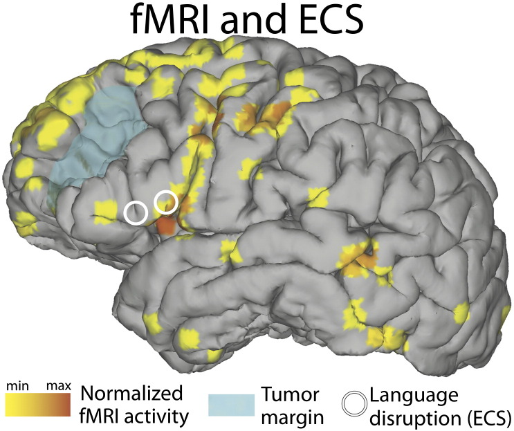
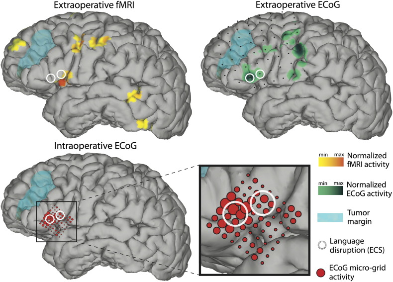
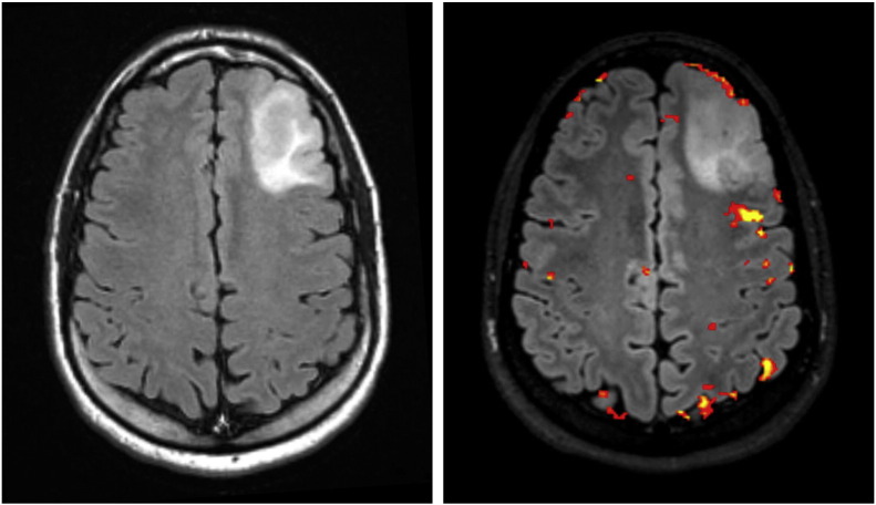
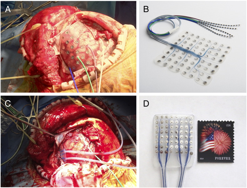
In this case report, we investigated the utility and practicality of passive intraoperative functional mapping of expressive language cortex using high-resolution electrocorticography (ECoG). The patient presented here experienced new-onset seizures caused by a medium-grade tumor in very close proximity to expressive language regions. In preparation of tumor resection, the patient underwent multiple functional language mapping procedures. We examined the relationship of results obtained with intraoperative high-resolution ECoG, extraoperative ECoG utilizing a conventional subdural grid, extraoperative electrical cortical stimulation (ECS) mapping, and functional magnetic resonance imaging (fMRI). Our results demonstrate that intraoperative mapping using high-resolution ECoG is feasible and, within minutes, produces results that are qualitatively concordant to those achieved by extraoperative mapping modalities. They also suggest that functional language mapping of expressive language areas with ECoG may prove useful in many intraoperative conditions given its time efficiency and safety. Finally, they demonstrate that integration of results from multiple functional mapping techniques, both intraoperative and extraoperative, may serve to improve the confidence in or precision of functional localization when pathology encroaches upon eloquent language cortex.
在本病例报告中,我们研究了使用高分辨率脑皮层电图(ECoG)对表达性语言皮层进行术中被动功能图谱绘制的实用性和可行性。此处报告的患者因靠近表达性语言区域的中级肿瘤引发了新发癫痫。在准备进行肿瘤切除时,患者接受了多次功能性语言图谱绘制程序。我们检查了术中高分辨率ECoG、使用传统硬膜下网格的术后ECoG、术后皮层电刺激(ECS)图谱绘制以及功能磁共振成像(fMRI)所获得结果之间的关系。我们的结果表明,使用高分辨率ECoG进行术中图谱绘制是可行的,并且在数分钟内即可产生与术后图谱绘制方式所获得的结果在质量上相符的结果。这些结果还表明,鉴于其时间效率和安全性,利用ECoG对表达性语言区域进行功能性语言图谱绘制在许多术中情况下可能会被证明是有用的。最后,这些结果表明,当病变侵犯明确的语言皮层时,整合术中及术后多种功能性图谱绘制技术的结果可能有助于提高功能定位的可信度或精度。