Carvalho Thiago Saads, Baumann Tommy, Lussi Adrian
Department of Preventive, Restorative and Pediatric Dentistry, University of Bern, Freiburgstrasse 7, CH-3010, Bern, Switzerland.
BMC Oral Health. 2016 Jul 7;17(1):14. doi: 10.1186/s12903-016-0231-y.
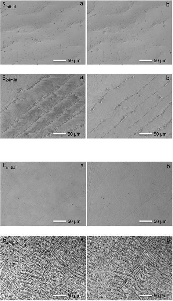
Erosive tooth wear (ETW) is clinically characterized by a loss of tooth surface, and different enamel depths may have different susceptibility to demineralization. Therefore, the aim of this in vitro pilot study was to assess if the progression of erosive demineralization is faster on teeth already presenting signs of ETW when compared to originally sound teeth.
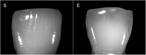
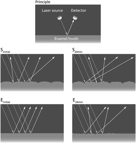
We selected 23 central incisors: 14 were clinically sound (Sound) and 9 presented clinical signs of early erosive tooth wear (ETW-teeth). The teeth were embedded in resin, leaving an uncovered window of native enamel (6.69 ± 2.30 mm(2)) on the incisal half of the labial surface. We measured enamel surface reflection intensity (SRI) initially and after each consecutive erosive challenge (1 % citric acid, total of 4, 8, 12, 16, 20 and 24 min). Calcium released to the citric acid was measured with an atomic absorption spectrometer.
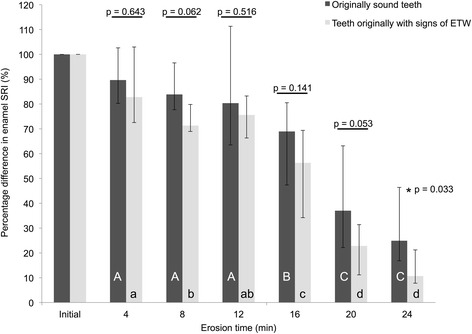
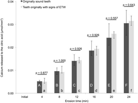
We observed higher initial SRI values in ETW-teeth than in Sound teeth (p = 0.007). During in vitro erosive demineralization, we observed that erosion on originally Sound teeth progressed significantly slower (p = 0.033) than on ETW-teeth: SRI decreased by 75 % (from 100 to 25 %) on Sound teeth, and by 89 % (from 100 to 11 %) on ETW-teeth. Calcium release increased during erosion, but presented no significant differences (p = 0.643) between originally Sound (0.031 μmol/mm(2)) and ETW-teeth (0.032 μmol/mm(2)). There was satisfactory correlation between calcium release and rSRI values (r s = -0.66).
The optical reflectometer distinguished originally sound teeth from those with signs of ETW, and the results suggest that acid demineralization progresses differently on teeth already presenting clinical signs of ETW than on sound teeth.
侵蚀性牙磨损(ETW)的临床特征为牙面丧失,且不同的釉质深度对脱矿的敏感性可能不同。因此,这项体外初步研究的目的是评估与原本健康的牙齿相比,已经出现ETW迹象的牙齿上侵蚀性脱矿的进展是否更快。
我们选取了23颗中切牙:14颗临床健康(健康组),9颗呈现早期侵蚀性牙磨损的临床迹象(ETW牙齿组)。将牙齿嵌入树脂中,在唇面切缘一半处留出未覆盖的天然釉质窗口(6.69±2.30平方毫米)。我们在初始时以及每次连续侵蚀挑战(1%柠檬酸,共4、8、12、16、20和24分钟)后测量釉质表面反射强度(SRI)。用原子吸收光谱仪测量释放到柠檬酸中的钙。
我们观察到ETW牙齿组的初始SRI值高于健康组牙齿(p = 0.007)。在体外侵蚀性脱矿过程中,我们观察到原本健康的牙齿上的侵蚀进展明显慢于ETW牙齿组(p = 0.033):健康组牙齿的SRI下降了75%(从100降至25%),而ETW牙齿组下降了89%(从100降至11%)。侵蚀过程中钙释放增加,但原本健康的牙齿(0.031微摩尔/平方毫米)和ETW牙齿组(0.032微摩尔/平方毫米)之间没有显著差异(p = 0.643)。钙释放与相对SRI值之间存在良好的相关性(rs = -0.66)。
光学反射仪能够区分原本健康的牙齿和有ETW迹象的牙齿,结果表明,酸脱矿在已经出现ETW临床迹象的牙齿上的进展与在健康牙齿上不同。