Latvala Siiri, Hedberg Jonas, Di Bucchianico Sebastiano, Möller Lennart, Odnevall Wallinder Inger, Elihn Karine, Karlsson Hanna L
Department of Environmental Science and Analytical Chemistry, Stockholm University, Stockholm, Sweden.
KTH Royal Institute of Technology, Division of Surface and Corrosion Science, School of Chemical Science and Engineering, Stockholm, Sweden.
PLoS One. 2016 Jul 19;11(7):e0159684. doi: 10.1371/journal.pone.0159684. eCollection 2016.
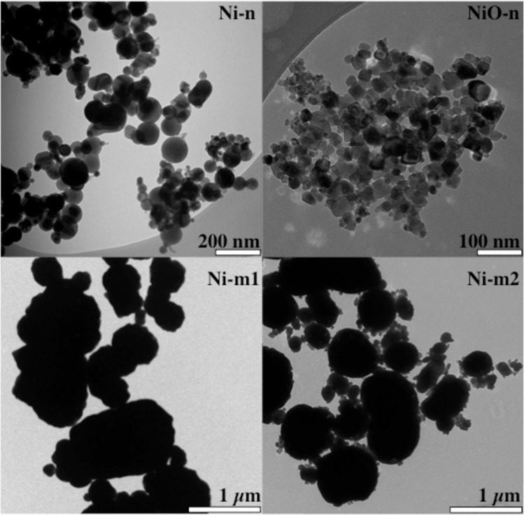
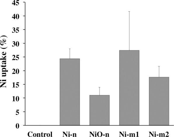
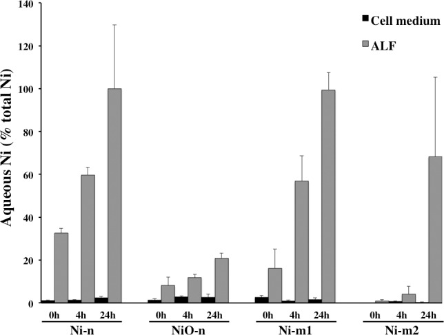
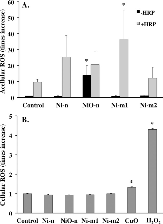
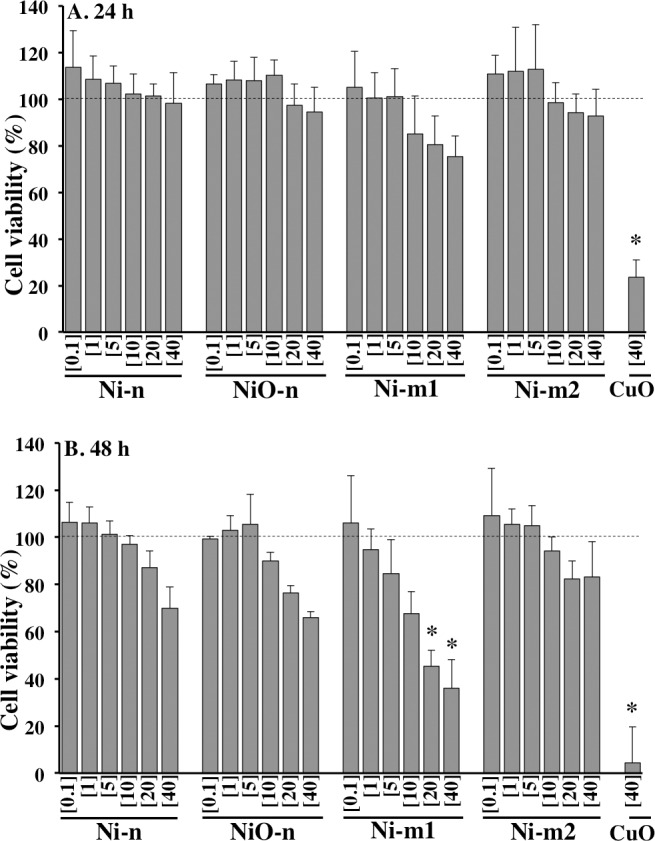
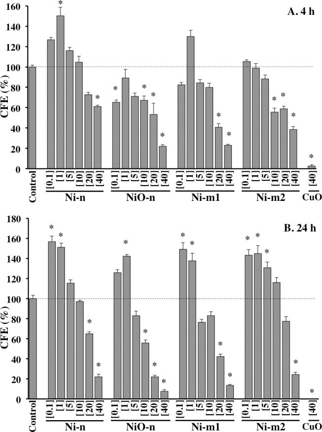
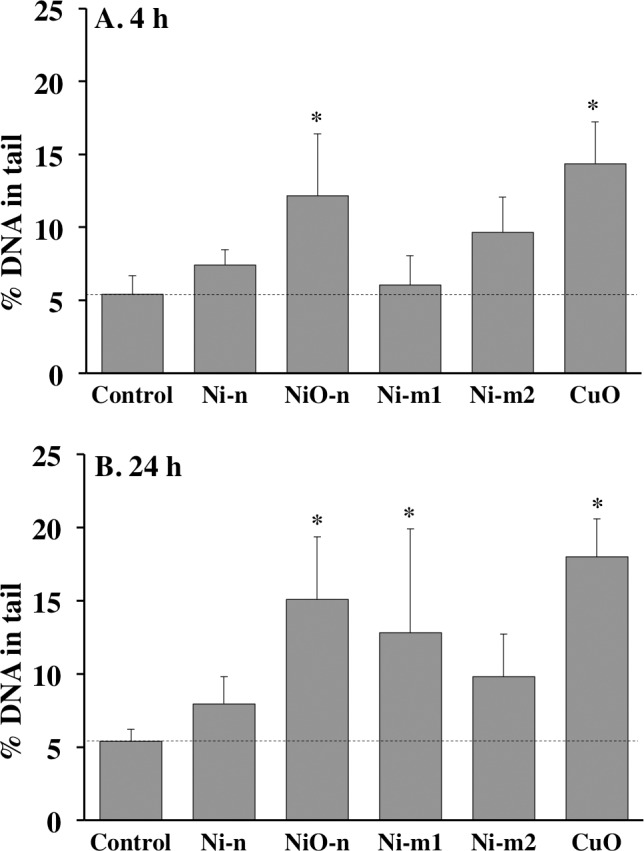
Occupational exposure to airborne nickel is associated with an elevated risk for respiratory tract diseases including lung cancer. Therefore, the increased production of Ni-containing nanoparticles necessitates a thorough assessment of their physical, chemical, as well as toxicological properties. The aim of this study was to investigate and compare the characteristics of nickel metal (Ni) and nickel oxide (NiO) particles with a focus on Ni release, reactive oxygen species (ROS) generation, cellular uptake, cytotoxicity and genotoxicity. Four Ni-containing particles of both nano-size (Ni-n and NiO-n) and micron-size (Ni-m1 and Ni-m2) were tested. The released amount of Ni in solution was notably higher in artificial lysosomal fluid (e.g. 80-100 wt% for metallic Ni) than in cell medium after 24h (ca. 1-3 wt% for all particles). Each of the particles was taken up by the cells within 4 h and they remained in the cells to a high extent after 24 h post-incubation. Thus, the high dissolution in ALF appeared not to reflect the particle dissolution in the cells. Ni-m1 showed the most pronounced effect on cell viability after 48 h (alamar blue assay) whereas all particles showed increased cytotoxicity in the highest doses (20-40 μg cm2) when assessed by colony forming efficiency (CFE). Interestingly an increased CFE, suggesting higher proliferation, was observed for all particles in low doses (0.1 or 1 μg cm-2). Ni-m1 and NiO-n were the most potent in causing acellular ROS and DNA damage. However, no intracellular ROS was detected for any of the particles. Taken together, micron-sized Ni (Ni-m1) was more reactive and toxic compared to the nano-sized Ni. Furthermore, this study underlines that the low dose effect in terms of increased proliferation observed for all particles should be further investigated in future studies.
职业性接触空气中的镍会增加患包括肺癌在内的呼吸道疾病的风险。因此,含镍纳米颗粒产量的增加需要对其物理、化学以及毒理学特性进行全面评估。本研究的目的是调查和比较镍金属(Ni)和氧化镍(NiO)颗粒的特性,重点关注镍的释放、活性氧(ROS)的产生、细胞摄取、细胞毒性和遗传毒性。测试了四种纳米尺寸(Ni-n和NiO-n)和微米尺寸(Ni-m1和Ni-m2)的含镍颗粒。24小时后,人工溶酶体液中镍的释放量(例如金属镍为80-100 wt%)明显高于细胞培养基中的释放量(所有颗粒约为1-3 wt%)。每种颗粒在4小时内被细胞摄取,孵育24小时后它们在细胞内仍有很高的留存率。因此,在人工溶酶体液中的高溶解性似乎并不能反映颗粒在细胞中的溶解情况。48小时后(alamar蓝检测法),Ni-m1对细胞活力的影响最为明显,而通过集落形成效率(CFE)评估时,所有颗粒在最高剂量(20-40 μg cm2)下均表现出细胞毒性增加。有趣的是,在低剂量(0.1或1 μg cm-2)下,所有颗粒的CFE均增加,表明增殖更高。Ni-m1和NiO-n在引起无细胞ROS和DNA损伤方面最为有效。然而,未检测到任何颗粒产生细胞内ROS。综上所述,微米尺寸的镍(Ni-m1)比纳米尺寸的镍更具反应性和毒性。此外,本研究强调,所有颗粒在增殖增加方面的低剂量效应应在未来的研究中进一步调查。