Gliga Anda R, Skoglund Sara, Wallinder Inger Odnevall, Fadeel Bengt, Karlsson Hanna L
Division of Molecular Toxicology, Institute of Environmental Medicine, Karolinska Institutet, SE-171 77 Stockholm, Sweden.
Part Fibre Toxicol. 2014 Feb 17;11:11. doi: 10.1186/1743-8977-11-11.
Silver nanoparticles (AgNPs) are currently one of the most manufactured nanomaterials. A wide range of toxicity studies have been performed on various AgNPs, but these studies report a high variation in toxicity and often lack proper particle characterization. The aim of this study was to investigate size- and coating-dependent toxicity of thoroughly characterized AgNPs following exposure of human lung cells and to explore the mechanisms of toxicity.
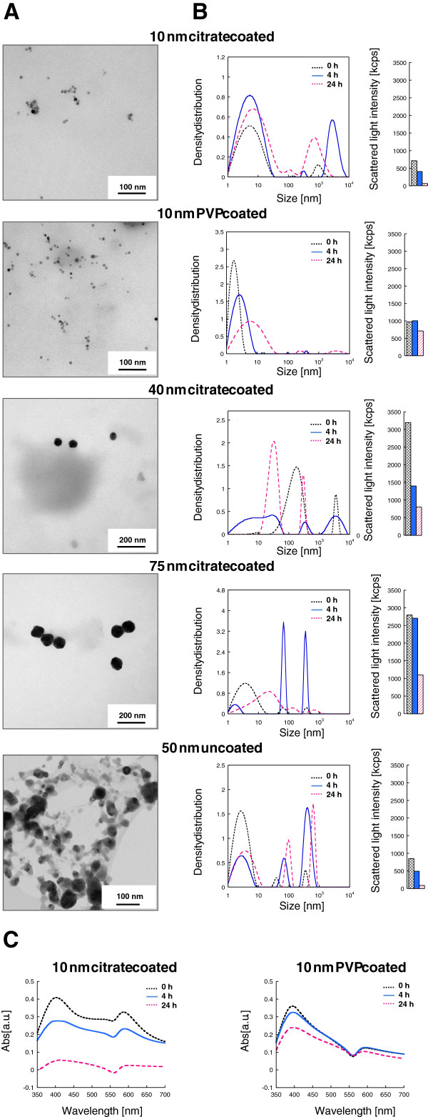
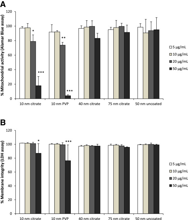
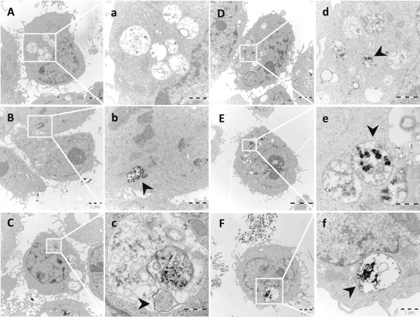
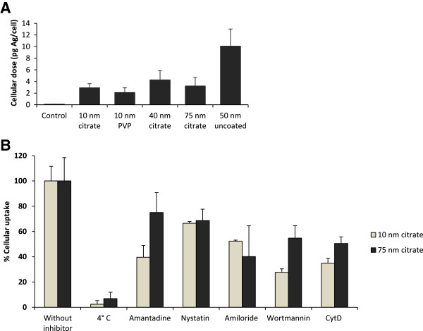
BEAS-2B cells were exposed to citrate coated AgNPs of different primary particle sizes (10, 40 and 75 nm) as well as to 10 nm PVP coated and 50 nm uncoated AgNPs. The particle agglomeration in cell medium was investigated by photon cross correlation spectroscopy (PCCS); cell viability by LDH and Alamar Blue assay; ROS induction by DCFH-DA assay; genotoxicity by alkaline comet assay and γH2AX foci formation; uptake and intracellular localization by transmission electron microscopy (TEM); and cellular dose as well as Ag release by atomic absorption spectroscopy (AAS).
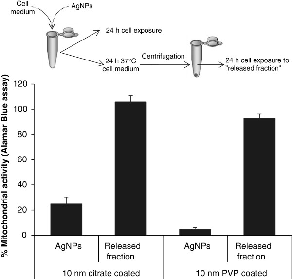
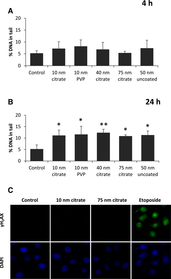
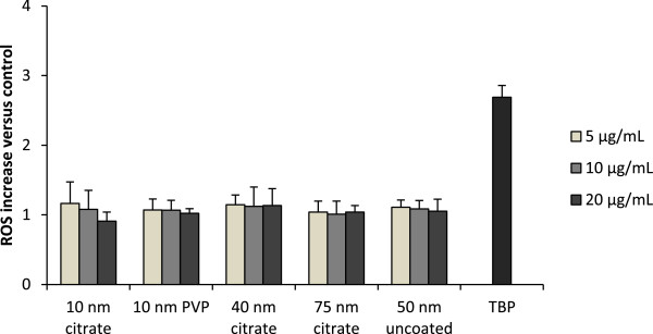
The results showed cytotoxicity only of the 10 nm particles independent of surface coating. In contrast, all AgNPs tested caused an increase in overall DNA damage after 24 h assessed by the comet assay, suggesting independent mechanisms for cytotoxicity and DNA damage. However, there was no γH2AX foci formation and no increased production of intracellular reactive oxygen species (ROS). The reasons for the higher toxicity of the 10 nm particles were explored by investigating particle agglomeration in cell medium, cellular uptake, intracellular localization and Ag release. Despite different agglomeration patterns, there was no evident difference in the uptake or intracellular localization of the citrate and PVP coated AgNPs. However, the 10 nm particles released significantly more Ag compared with all other AgNPs (approx. 24 wt% vs. 4-7 wt%) following 24 h in cell medium. The released fraction in cell medium did not induce any cytotoxicity, thus implying that intracellular Ag release was responsible for the toxicity.
This study shows that small AgNPs (10 nm) are cytotoxic for human lung cells and that the toxicity observed is associated with the rate of intracellular Ag release, a 'Trojan horse' effect.
银纳米颗粒(AgNPs)是目前生产最多的纳米材料之一。已对各种AgNPs进行了广泛的毒性研究,但这些研究报告的毒性差异很大,且常常缺乏适当的颗粒表征。本研究的目的是调查经过充分表征的AgNPs在暴露于人类肺细胞后其大小和涂层依赖性毒性,并探索毒性机制。
将BEAS-2B细胞暴露于不同初级粒径(10、40和75nm)的柠檬酸盐包被的AgNPs,以及10nm聚乙烯吡咯烷酮(PVP)包被的和50nm未包被的AgNPs。通过光子交叉相关光谱法(PCCS)研究细胞培养基中的颗粒团聚;通过乳酸脱氢酶(LDH)和alamar蓝测定法研究细胞活力;通过2',7'-二氯二氢荧光素二乙酸酯(DCFH-DA)测定法研究活性氧(ROS)诱导;通过碱性彗星试验和γH2AX焦点形成研究遗传毒性;通过透射电子显微镜(TEM)研究摄取和细胞内定位;通过原子吸收光谱法(AAS)研究细胞剂量以及银释放。
结果显示仅10nm颗粒具有细胞毒性,与表面涂层无关。相比之下,通过彗星试验评估,所有测试的AgNPs在24小时后均导致总体DNA损伤增加,这表明细胞毒性和DNA损伤的机制相互独立。然而,没有γH2AX焦点形成,细胞内活性氧(ROS)的产生也没有增加。通过研究细胞培养基中的颗粒团聚、细胞摄取、细胞内定位和银释放,探索了10nm颗粒毒性更高的原因。尽管团聚模式不同,但柠檬酸盐和PVP包被的AgNPs在摄取或细胞内定位方面没有明显差异。然而,在细胞培养基中培养24小时后,10nm颗粒释放的银比所有其他AgNPs显著更多(约24重量%对4-7重量%)。细胞培养基中释放的部分没有诱导任何细胞毒性,因此表明细胞内银释放是毒性的原因。
本研究表明,小的AgNPs(10nm)对人类肺细胞具有细胞毒性,观察到的毒性与细胞内银释放速率有关,即“特洛伊木马”效应。