Guensch Dominik P, Nadeshalingam Gobinath, Fischer Kady, Stalder Aurelien F, Friedrich Matthias G
Philippa & Marvin Carsley CMR Centre at the Montreal Heart Institute, Montreal, QC, Canada.
Department of Anesthesiology and Pain Therapy, Inselspital, Bern University Hospital, University of Bern, Freiburgstrasse, 3010, Bern, Switzerland.
J Cardiovasc Magn Reson. 2016 Jul 20;18(1):42. doi: 10.1186/s12968-016-0262-1.
Oxygenation-sensitive (OS) Cardiovascular Magnetic Resonance (CMR) is a promising utility in the diagnosis of heart disease. Contrast in OS-CMR images is generated through deoxyhemoglobin in the tissue, which is negatively correlated with the signal intensity (SI). Thus, changing hematocrit levels may be a confounder in the interpretation of OS-CMR results. We hypothesized that hemodilution confounds the observed signal intensity in OS-CMR images.
Venous and arterial blood from five pigs was diluted with lactated Ringer solution in 10 % increments to 50 %. The changes in signal intensity (SI) were compared to changes in blood gases and hemoglobin concentration. We performed an OS-CMR scan in 21 healthy volunteers using vasoactive breathing stimuli at baseline, which was then repeated after rapid infusion of 1 L of lactated Ringer's solution within 5-8 min. Changes of SI were measured and compared between the hydration states.
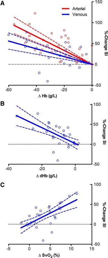
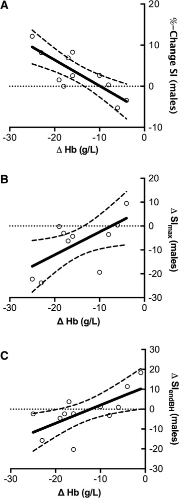
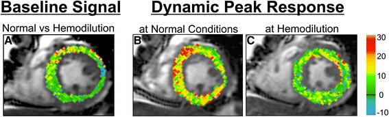
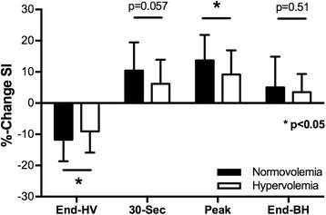
The % change in SI from baseline for arterial (r = -0.67, p < 0.0001) and venous blood (r = -0.55, p = 0.002) were negatively correlated with the changes in hemoglobin (Hb). SI changes in venous blood were also associated with SO2 (r = 0.68, p < 0.0001) and deoxyHb concentration (-0.65, p < 0.0001). In healthy volunteers, rapid infusion resulted in a significant drop in the hemoglobin concentration (142.5 ± 15.2 g/L vs. 128.8 ± 15.2 g/L; p < 0.0001). Baseline myocardial SI increased by 3.0 ± 5.7 % (p = 0.026) following rapid infusion, and in males there was a strong association between the change in hemoglobin concentration and % changes in SI (r = 0.82, p = 0.002). After hyperhydration, the SI response after hyperventilation was attenuated (HV, p = 0.037), as was the maximum SI increase during apnea (p = 0.012). The extent of SI attenuation was correlated with the reduction in hemoglobin concentration at the end of apnea (r = 0.55, p = 0.012) for all subjects and at maximal SI (r = 0.63, p = 0.037) and the end of breath-hold (r = 0.68, p = 0.016) for males only.
In dynamic studies using oxygenation-sensitive CMR, the hematocrit level affects baseline signal intensity and the observed signal intensity response. Thus, the hydration status of the patient may be a confounder for OS-CMR image analysis.
氧敏型(OS)心血管磁共振成像(CMR)在心脏病诊断中具有广阔前景。OS-CMR图像中的对比度由组织中的脱氧血红蛋白产生,其与信号强度(SI)呈负相关。因此,血细胞比容水平的变化可能会干扰OS-CMR结果的解读。我们推测血液稀释会干扰OS-CMR图像中观察到的信号强度。
将五只猪的静脉血和动脉血用乳酸林格氏液以10%的增量稀释至50%。将信号强度(SI)的变化与血气和血红蛋白浓度的变化进行比较。我们对21名健康志愿者进行了OS-CMR扫描,在基线时使用血管活性呼吸刺激,然后在5 - 8分钟内快速输注1升乳酸林格氏液后重复扫描。测量并比较不同水化状态下的SI变化。
动脉血(r = -0.67,p < 0.0001)和静脉血(r = -0.55,p = 0.002)相对于基线的SI变化百分比与血红蛋白(Hb)的变化呈负相关。静脉血中的SI变化也与SO2(r = 0.68,p < 0.0001)和脱氧血红蛋白浓度(-0.65,p < 0.0001)相关。在健康志愿者中,快速输注导致血红蛋白浓度显著下降(142.5 ± 15.2 g/L vs. 128.8 ± 15.2 g/L;p < 0.0001)。快速输注后,基线心肌SI增加了3.0 ± 5.7%(p = 0.026),并且在男性中,血红蛋白浓度的变化与SI变化百分比之间存在很强的相关性(r = 0.82,p = 0.002)。补液后,过度通气后的SI反应减弱(HV,p = 0.037),呼吸暂停期间SI的最大增加也减弱(p = 0.012)。所有受试者的SI衰减程度与呼吸暂停结束时血红蛋白浓度的降低相关(r = 0.55,p = 0.012),仅男性在最大SI时(r = 0.63,p = 0.037)和屏气结束时(r = 0.68,p = 0.016)相关。
在使用氧敏型CMR的动态研究中,血细胞比容水平会影响基线信号强度和观察到的信号强度反应。因此,患者的水化状态可能是OS-CMR图像分析的一个干扰因素。