Kim Won Young, Kim Jin Bum, Nam Taek Kyun, Kim Young Baeg, Park Seung Won
Department of Neurosurgery, Chung-Ang University College of Medicine, Seoul, Korea.
Korean J Spine. 2016 Jun;13(2):67-70. doi: 10.14245/kjs.2016.13.2.67. Epub 2016 Jun 30.
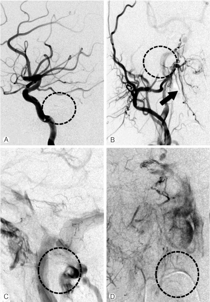
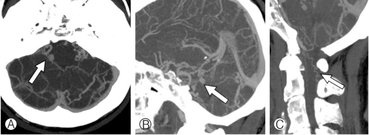
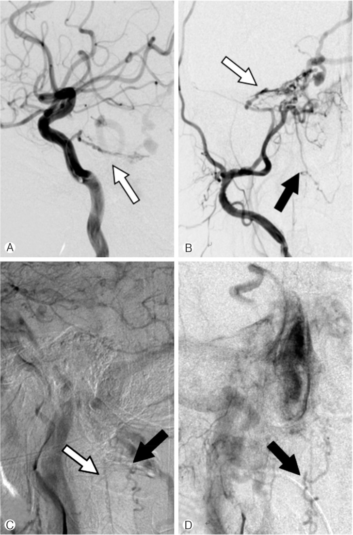
Intracranial dural arteriovenous fistula (dAVF) usually results in various problems in the brain. But it can be presented as a myelopathy, which may make early diagnosis and management to be difficult. We recently experienced a case of cervical myelopathy caused by intracranial dAVF. A 60-year-old man presented with a 3-year history of gait disturbance due to a progressive weakness of both legs. Neurological examination revealed spastic paraparesis (grade IV) and Babinski sign on both sides. Magnetic resonance imaging showed serpentine vascular signal voids at C2-T1 on T2-weighted image with increased signal intensity and swelling of spinal cord at C1-C4. We performed a brain computed tomography angiography and found intracranial dAVF with multiple arteriovenous shunts. Venous drainages were noted at tentorial veins and cervical perimedullary veins. After Onyx embolization, the patient showed gradual improvement in motor power and gait disturbance. The venous drainage pattern is a well-known prognostic factor of dAVF. In our case, the intracranial dAVF drained to spinal perimedullary vein, which seemed to result in the ischemic myelopathy. Although it is rare condition, it sometimes can cause serious complications. Therefore, we should keep in mind the possibility of intracranial dAVF when a patient presents myelopathy.
颅内硬脑膜动静脉瘘(dAVF)通常会导致大脑出现各种问题。但它也可能表现为脊髓病,这可能会使早期诊断和治疗变得困难。我们最近遇到了一例由颅内dAVF引起的颈髓病病例。一名60岁男性因双腿进行性无力出现了3年的步态障碍病史。神经系统检查发现双侧痉挛性截瘫(IV级)和巴宾斯基征。磁共振成像显示在T2加权图像上C2 - T1水平有蜿蜒的血管信号缺失,C1 - C4脊髓信号强度增加且肿胀。我们进行了脑部计算机断层血管造影,发现颅内dAVF伴有多处动静脉分流。观察到静脉引流至小脑幕静脉和颈髓周静脉。经Onyx栓塞治疗后,患者的运动能力和步态障碍逐渐改善。静脉引流模式是dAVF一个众所周知的预后因素。在我们的病例中,颅内dAVF引流至脊髓周静脉,这似乎导致了缺血性脊髓病。虽然这种情况罕见,但有时会引起严重并发症。因此,当患者出现脊髓病时,我们应牢记颅内dAVF的可能性。