Miyata Makoto, Hamaguchi Tasuku
Department of Biology, Graduate School of Science, Osaka City UniversityOsaka, Japan; The OCU Advanced Research Institute for Natural Science and Technology, Osaka City UniversityOsaka, Japan.
Front Microbiol. 2016 Jun 28;7:960. doi: 10.3389/fmicb.2016.00960. eCollection 2016.
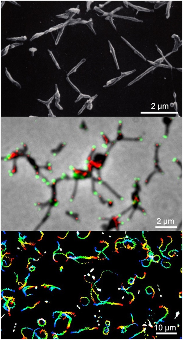
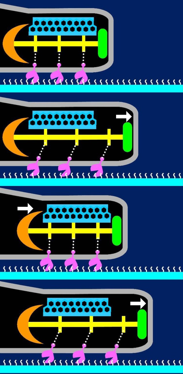
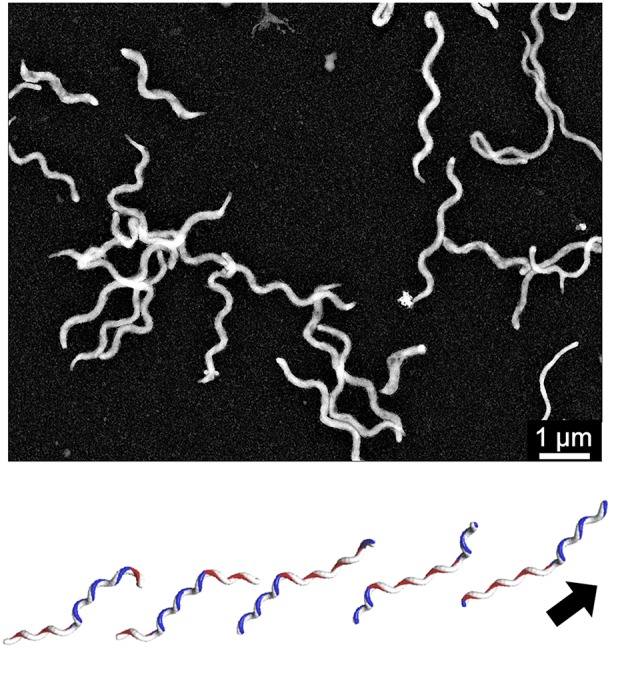
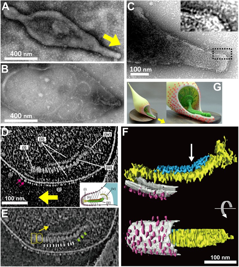
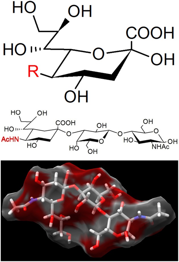
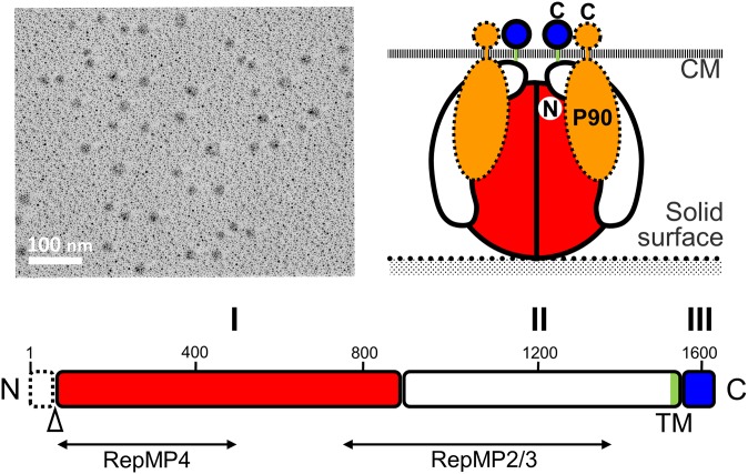
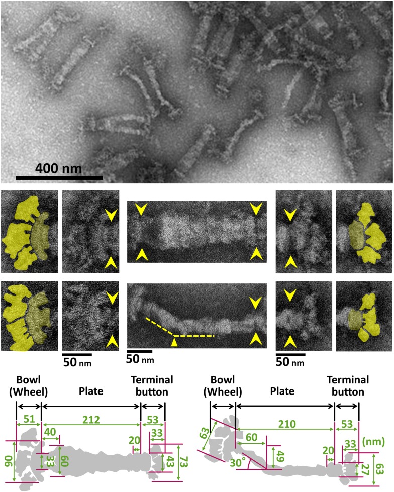
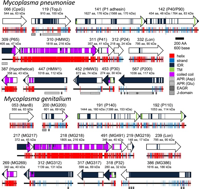
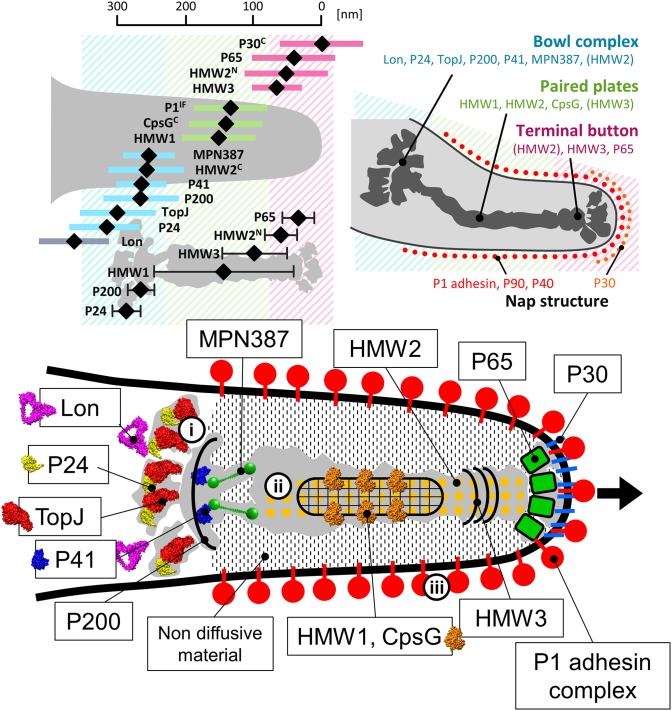
Mycoplasma pneumoniae forms a membrane protrusion at a cell pole and is known to adhere to solid surfaces, including animal cells, and can glide on these surfaces with a speed up to 1 μm per second. Notably, gliding appears to be involved in the infectious process in addition to providing the bacteria with a means of escaping the host's immune systems. However, the genome of M. pneumoniae does not encode any of the known genes found in other bacterial motility systems or any conventional motor proteins that are responsible for eukaryotic motility. Thus, further analysis of the mechanism underlying M. pneumoniae gliding is warranted. The gliding machinery formed as the membrane protrusion can be divided into the surface and internal structures. On the surface, P1 adhesin, a 170 kDa transmembrane protein forms an adhesin complex with other two proteins. The internal structure features a terminal button, paired plates, and a bowl (wheel) complex. In total, the organelle is composed of more than 15 proteins. By integrating the currently available information by genetics, microscopy, and structural analyses, we have suggested a working model for the architecture of the organelle. Furthermore, in this article, we suggest and discuss a possible mechanism of gliding based on the structural model, in which the force generated around the bowl complex transmits through the paired plates, reaching the adhesin complex, resulting in the repeated catch of sialylated oligosaccharides on the host surface by the adhesin complex.
肺炎支原体在细胞极形成膜突出物,已知其可附着于包括动物细胞在内的固体表面,并能以高达每秒1微米的速度在这些表面上滑行。值得注意的是,滑行似乎不仅为细菌提供了逃避宿主免疫系统的手段,还参与了感染过程。然而,肺炎支原体的基因组并未编码其他细菌运动系统中发现的任何已知基因,也没有编码负责真核生物运动的任何传统运动蛋白。因此,有必要进一步分析肺炎支原体滑行的潜在机制。作为膜突出物形成的滑行机制可分为表面结构和内部结构。在表面,P1黏附素是一种170 kDa的跨膜蛋白,与其他两种蛋白形成黏附素复合物。内部结构的特征是末端纽扣、成对的板和碗(轮)状复合物。该细胞器总共由超过15种蛋白质组成。通过整合遗传学、显微镜学和结构分析等当前可用信息,我们提出了该细胞器结构的工作模型。此外,在本文中,我们基于结构模型提出并讨论了一种可能的滑行机制,即碗状复合物周围产生的力通过成对的板传递,到达黏附素复合物,导致黏附素复合物反复捕获宿主表面的唾液酸化寡糖。