Kawamoto Akihiro, Matsuo Lisa, Kato Takayuki, Yamamoto Hiroki, Namba Keiichi, Miyata Makoto
Graduate School of Frontier Biosciences, Osaka University, Suita, Osaka, Japan.
Department of Biology, Graduate School of Science, Osaka City University, Sumiyoshi-ku, Osaka, Japan.
mBio. 2016 Apr 12;7(2):e00243-16. doi: 10.1128/mBio.00243-16.
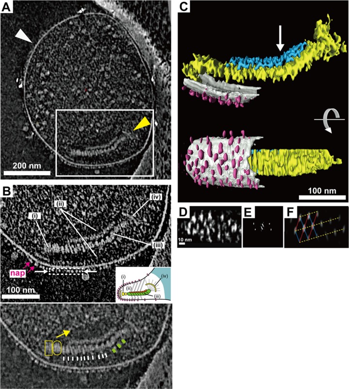
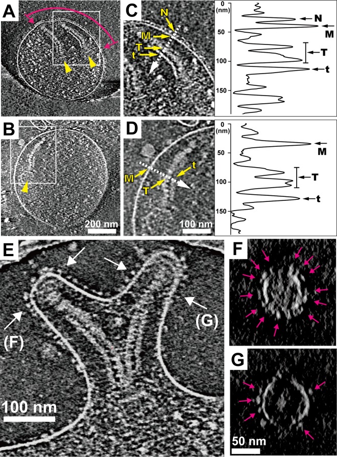
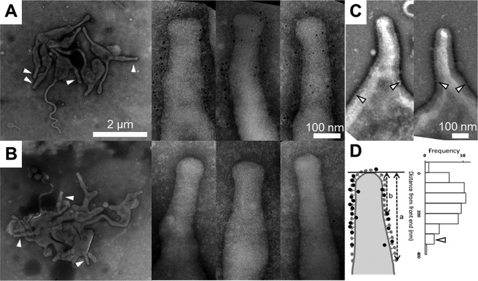
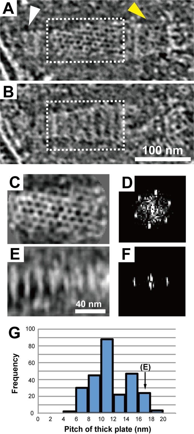
Mycoplasma pneumoniae, a pathogenic bacterium, glides on host surfaces using a unique mechanism. It forms an attachment organelle at a cell pole as a protrusion comprised of knoblike surface structures and an internal core. Here, we analyzed the three-dimensional structure of the organelle in detail by electron cryotomography. On the surface, knoblike particles formed a two-dimensional array, albeit with limited regularity. Analyses using a nonbinding mutant and an antibody showed that the knoblike particles correspond to a naplike structure that has been observed by negative-staining electron microscopy and is likely to be formed as a complex of P1 adhesin, the key protein for binding and gliding. The paired thin and thick plates feature a rigid hexagonal lattice and striations with highly variable repeat distances, respectively. The combination of variable and invariant structures in the internal core and the P1 adhesin array on the surface suggest a model in which axial extension and compression of the thick plate along a rigid thin plate is coupled with attachment to and detachment from the substrate during gliding.
Human mycoplasma pneumonia, epidemic all over the world in recent years, is caused by a pathogenic bacterium,Mycoplasma pneumoniae This tiny bacterium, about 2 µm in cell body length, glides on the surface of the human trachea to infect the host by binding to sialylated oligosaccharides, which are also the binding targets of influenza viruses. The mechanism of mycoplasmal gliding motility is not related to any other well-studied motility systems, such as bacterial flagella and cytoplasmic motor proteins. Here, we visualized the attachment organelle, a cellular architecture for gliding, three dimensionally by using electron cryotomography and other conventional methods. A possible gliding mechanism has been suggested based on the architectural images.
肺炎支原体是一种病原菌,它利用独特的机制在宿主表面滑行。它在细胞极形成一个附着细胞器,作为一个由球状表面结构和内部核心组成的突起。在这里,我们通过电子冷冻断层扫描详细分析了该细胞器的三维结构。在表面,球状颗粒形成二维阵列,尽管规律性有限。使用非结合突变体和抗体进行的分析表明,球状颗粒对应于通过负染色电子显微镜观察到的绒毛状结构,并且可能是由P1黏附素(结合和滑行的关键蛋白)形成的复合物。成对的薄板和厚板分别具有刚性六边形晶格和重复距离高度可变的条纹。内部核心中可变和不变结构与表面P1黏附素阵列的组合提示了一种模型,即厚板沿刚性薄板的轴向伸展和压缩与滑行过程中与底物的附着和脱离相耦合。
近年来在全球流行的人类支原体肺炎是由病原菌肺炎支原体引起的。这种微小的细菌,细胞体长约2微米,在人类气管表面滑行,通过与唾液酸化寡糖结合来感染宿主,而唾液酸化寡糖也是流感病毒的结合靶点。支原体滑行运动的机制与任何其他经过充分研究的运动系统无关,如细菌鞭毛和细胞质运动蛋白。在这里,我们使用电子冷冻断层扫描和其他传统方法对附着细胞器(一种用于滑行的细胞结构)进行了三维可视化。基于这些结构图像提出了一种可能的滑行机制。