Sudo Haruka, Nakajima Kazunori
Department of Biochemistry, The Nippon Dental University School of Life Dentistry at Tokyo, Chiyoda-ku, Tokyo 102-8159, Japan.
Department of Anatomy, Keio University School of Medicine, Shinjuku-ku, Tokyo 160-8582, Japan.
Oncotarget. 2016 Aug 16;7(33):53712-53734. doi: 10.18632/oncotarget.10728.
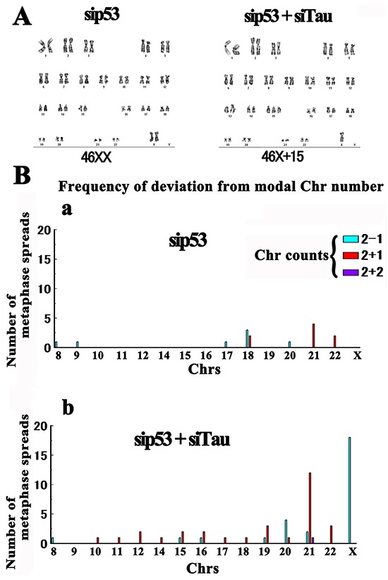
The microtubule associated-protein tau has been identified as an effective positive prognostic indicator in breast cancer. To explore the physiological function of tau in early carcinogenesis, endogenous tau was knocked down in primary cultured human mammary epithelial cells. This resulted in chromosome-bridging during anaphase followed by micronucleation, both of which were suppressed by a further katanin-like1 knockdown. We also detected that the exogenously expressed katanin-like1 induction of cellular transformation is prevented by exogenous tau in rat fibroblasts. The mutant katanin-like1 (L123V) identified in breast cancer showed an increase in this transformation capacity as well as microtubule severing activity resistant to tau. The tau knockdown resulted in a loss of the kinetochore fibers on which tau is normally localized. This physical fragility was also observed in isolated tau-knockdown mitotic spindles, supporting the relevance of microtubule damage to the onset of transformation. The karyotyping of tau-knockdown cells showed increased frequency of loss of one X chromosome, further suggesting the involvement of tau in breast tumorigenesis. We propose that tau may contribute to tumor progression by protecting spindle microtubules from excess severing by katanin-like1. We also present data indicating that the microtubule-binding octapeptide NAP is a candidate modifier against the tau deficiency in tumor cells.
微管相关蛋白tau已被确定为乳腺癌中一种有效的阳性预后指标。为了探究tau在早期致癌过程中的生理功能,在原代培养的人乳腺上皮细胞中敲低内源性tau。这导致后期出现染色体桥接,随后形成微核,而进一步敲低katanin样蛋白1可抑制这两种现象。我们还检测到,在大鼠成纤维细胞中,外源性tau可阻止外源性表达的katanin样蛋白1诱导的细胞转化。在乳腺癌中鉴定出的突变型katanin样蛋白1(L123V)显示这种转化能力增强,以及对tau具有抗性的微管切断活性。tau敲低导致tau正常定位的动粒纤维丢失。在分离的tau敲低有丝分裂纺锤体中也观察到这种物理脆弱性,支持微管损伤与转化起始的相关性。tau敲低细胞的核型分析显示一条X染色体丢失的频率增加,进一步表明tau参与乳腺癌发生。我们提出,tau可能通过保护纺锤体微管免受katanin样蛋白1过度切断而促进肿瘤进展。我们还提供数据表明,微管结合八肽NAP是肿瘤细胞中tau缺乏的候选修饰剂。