Kang Daesung, Liu Yuelu, Miskovic Vladimir, Keil Andreas, Ding Mingzhou
The J. Crayton Pruitt Family Department of Biomedical Engineering, University of Florida, Gainesville, Florida, USA.
Department of Psychology and Center for Affective Science, State University of New York at Binghamton, Binghamton, New York, USA.
Psychophysiology. 2016 Nov;53(11):1627-1638. doi: 10.1111/psyp.12731. Epub 2016 Jul 25.
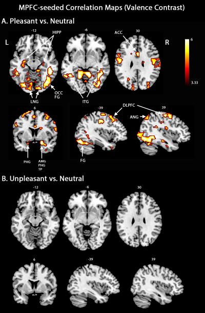
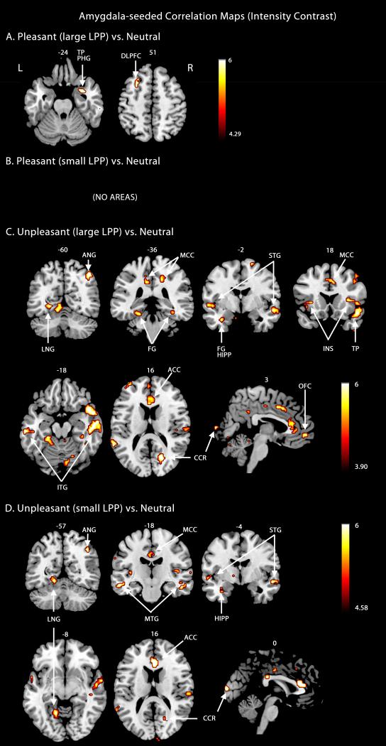
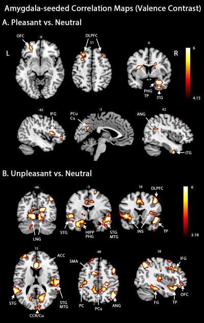
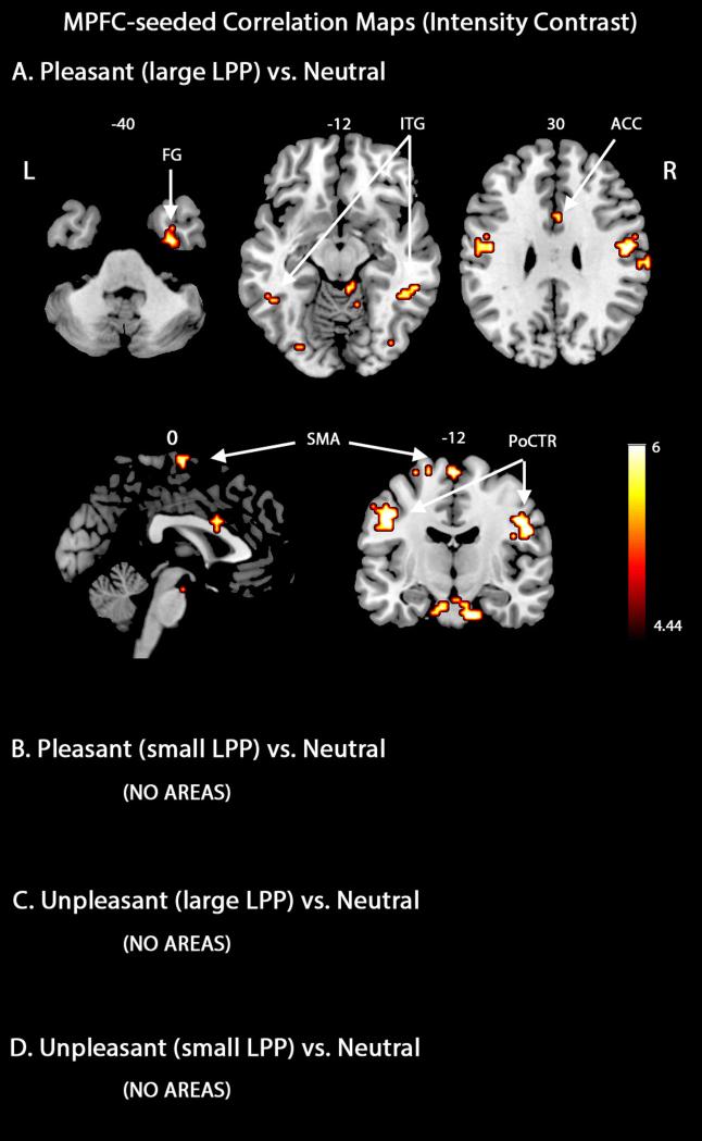
It has been hypothesized that the medial prefrontal cortex (mPFC) is a hub in the network that mediates appetitive responses whereas the amygdala is thought to mediate both aversive and appetitive processing. Both structures may facilitate adaptive responses to emotional challenge by linking perception, attention, memory, and motor circuits. We provide an initial exploration of these hypotheses by recording simultaneous EEG-fMRI in eleven participants viewing affective pictures. MPFC- and amygdala-seeded functional connectivity maps were generated by applying the beta-series correlation method. The mPFC-seeded correlation map encompassed visual regions, sensorimotor areas, prefrontal cortex, and medial temporal lobe structures, exclusively for pleasant content. For the amygdala-seeded correlation map, a similar set of distributed brain areas appeared in the unpleasant-neutral contrast, with the addition of structures such as the insula and thalamus. A substantially sparser network was recruited for the pleasant-neutral contrast. Using the late positive potential (LPP) to index the intensity of emotional engagement, functional connectivity was found to be stronger in trials with larger LPP. These results demonstrate that mPFC-mediated functional interactions are engaged specifically during appetitive processing, whereas the amygdala is coupled to distinct sets of brain regions during both aversive and appetitive processing. The strength of these interactions varies as a function of the intensity of emotional engagement.
有假说认为,内侧前额叶皮质(mPFC)是介导奖赏性反应的神经网络中的一个枢纽,而杏仁核则被认为介导厌恶和奖赏性加工。这两个结构可能通过连接感知、注意力、记忆和运动回路,促进对情绪挑战的适应性反应。我们通过记录11名观看情感图片的参与者的同步脑电图-功能磁共振成像,对这些假说进行了初步探索。通过应用β系列相关方法生成了以MPFC和杏仁核为种子的功能连接图谱。以mPFC为种子的相关图谱仅在愉悦内容的情况下,涵盖了视觉区域、感觉运动区域、前额叶皮质和内侧颞叶结构。对于以杏仁核为种子的相关图谱,在不愉快-中性对比中出现了一组类似的分布式脑区,还包括岛叶和丘脑等结构。在愉悦-中性对比中招募的网络要稀疏得多。使用晚期正电位(LPP)来索引情绪参与的强度,发现在LPP较大的试验中功能连接更强。这些结果表明,mPFC介导的功能相互作用在奖赏性加工过程中特异性地参与,而杏仁核在厌恶和奖赏性加工过程中都与不同的脑区集合相连。这些相互作用的强度随情绪参与强度而变化。