Leung J W, Sung J Y, Costerton J W
Department of Medicine, Chinese University of Hong Kong, Prince of Wales Hospital, Shatin, New Territories.
J Clin Microbiol. 1989 May;27(5):915-21. doi: 10.1128/jcm.27.5.915-921.1989.
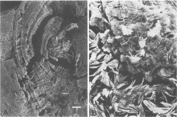
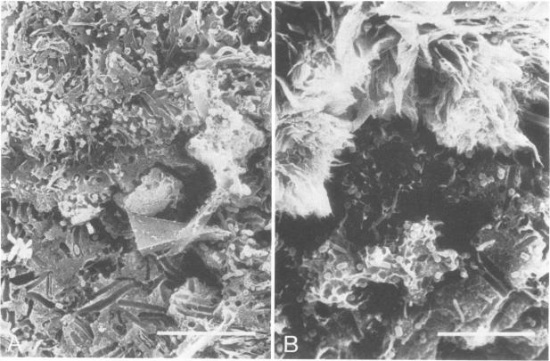
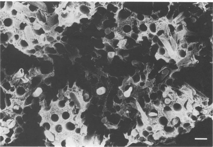
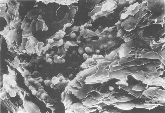
Bacteriological and morphological studies of 38 brown pigment common duct stones were performed. Stone cultures were positive for bacteria in 80.5% of those studied. Enterococci were the most common organisms that were isolated. Scanning electron microscopy showed the presence of bacteria in 84.2% of the stones. The bacteria were seen embedded within an amorphous matrix in alternating layers of flakelike crystals. Transmission electron microscopy showed the presence of gram-positive and gram-negative bacteria surrounded by a ruthenium red-stained exopolysaccharide material. Results of the bacteriological and morphological studies confirmed the close relationship between the presence of bacteria and the development of brown pigment stones.
对38颗棕色色素性胆总管结石进行了细菌学和形态学研究。在所研究的结石中,80.5%的结石培养出细菌。肠球菌是最常分离出的微生物。扫描电子显微镜显示84.2%的结石中有细菌存在。可见细菌包埋于片状晶体交替层的无定形基质中。透射电子显微镜显示革兰氏阳性菌和革兰氏阴性菌被钌红染色的胞外多糖物质包围。细菌学和形态学研究结果证实了细菌的存在与棕色色素结石形成之间的密切关系。