Reuter Björn, Venus Alexander, Heiler Patrick, Schad Lothar, Ebert Anne, Hennerici Michael G, Grudzenski Saskia, Fatar Marc
Department of Neurology and Neurophysiology, Freiburg University Freiburg, Germany.
Department of Neurology, Universitätsmedizin Mannheim, Heidelberg University Mannheim, Germany.
Front Aging Neurosci. 2016 Jul 8;8:170. doi: 10.3389/fnagi.2016.00170. eCollection 2016.
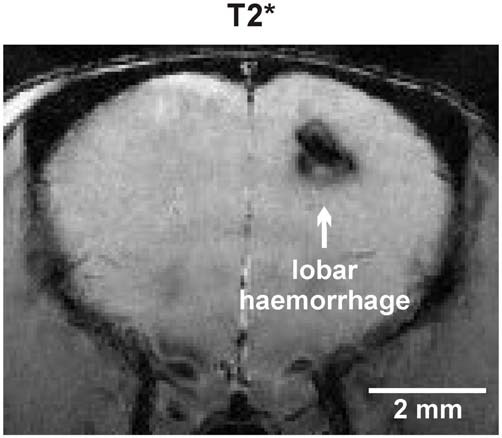
Cerebral amyloid angiopathy (CAA) is characterized by extracellular deposition of amyloid β (Aβ) around cerebral arteries and capillaries and leads to an increased risk for vascular dementia, spontaneous lobar hemorrhage, convexal subarachnoid hemorrhage, and transient focal neurological episodes, which might be an indicator of imminent spontaneous intracerebral hemorrhage. In CAA cerebral microbleeds (cMBs) with a cortical/juxtacortical distribution are frequently observed in standard magnetic resonance imaging (MRI). In vivo MRI of transgenic mouse models of CAA may serve as a useful tool to investigate translational aspects of the disease.
APP23-transgenic mice demonstrate cerebrovascular Aβ deposition with subsequent neuropathological changes characteristic for CAA. We performed a 9.4 Tesla high field MRI study using T2, T2* and time of flight-magnetic resonance angiograpy (TOF-MRA) sequences in APP23-transgenic mice and wildtype (wt) littermates at the age of 8, 12, 16, 20 and 24 months, respectively. Numbers, size, and location of cMBs are reported.
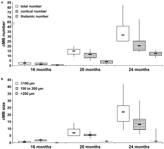
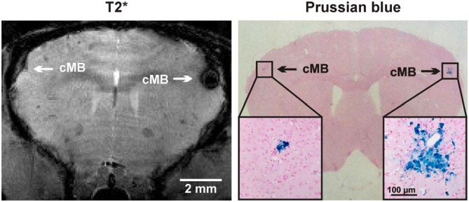
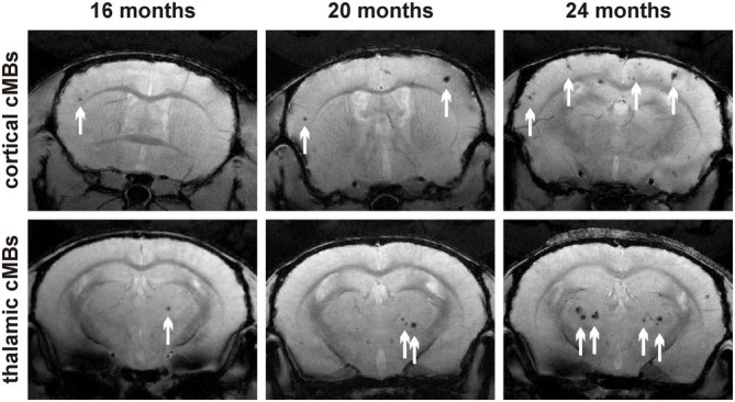
T2* imaging demonstrated cMBs (diameter 50-300 μm) located in the neocortex and, to a lesser degree, in the thalamus. cMBs were detected at the earliest at 16 months of age. Numbers increased exponentially with age, with 2.5 ± 2 (median ± interquartilrange) at 16 months, 15 ± 6 at 20 months, and 31.5 ± 17 at 24 months of age, respectively.
We report the temporal and spatial development of cMBs in the aging APP23-transgenic mouse model which develops characteristic pathological patterns known from human CAA. We expect this mouse model to serve as a useful tool to non-invasively monitor mid- and longterm translational aspects of CAA and to investigate experimental therapeutic strategies in longitudinal studies.
脑淀粉样血管病(CAA)的特征是淀粉样β蛋白(Aβ)在脑动脉和毛细血管周围细胞外沉积,并导致血管性痴呆、自发性脑叶出血、脑凸面蛛网膜下腔出血以及短暂性局灶性神经发作的风险增加,后者可能是即将发生自发性脑出血的一个指标。在CAA中,标准磁共振成像(MRI)经常观察到具有皮质/皮质旁分布的脑微出血(cMBs)。CAA转基因小鼠模型的体内MRI可能是研究该疾病转化方面的有用工具。
APP23转基因小鼠表现出脑血管Aβ沉积,并伴有CAA特有的后续神经病理变化。我们分别在8、12、16、20和24个月龄的APP23转基因小鼠和野生型(wt)同窝小鼠中,使用T2、T2*和时间飞跃磁共振血管造影(TOF-MRA)序列进行了一项9.4特斯拉高场MRI研究。报告了cMBs的数量、大小和位置。
T2*成像显示cMBs(直径50 - 300μm)位于新皮质,在较小程度上位于丘脑。最早在16个月龄时检测到cMBs。数量随年龄呈指数增加,16个月龄时为2.5±2(中位数±四分位间距),20个月龄时为15±6,24个月龄时为31.5±17。
我们报告了衰老的APP23转基因小鼠模型中cMBs的时间和空间发展情况,该模型呈现出人类CAA已知的特征性病理模式。我们期望这个小鼠模型能成为一个有用的工具,用于非侵入性监测CAA的中长期转化方面,并在纵向研究中研究实验性治疗策略。