Pires Flávio O, Dos Anjos Carlos A S, Covolan Roberto J M, Pinheiro Fabiano A, St Clair Gibson Alan, Noakes Timothy D, Magalhães Fernando H, Ugrinowitsch Carlos
Exercise Psychophysiology Research Group, School of Arts, Sciences, and Humanities, University of São PauloSão Paulo, Brazil; Department of Sport, School of Physical Education and Sport, University of São PauloSão Paulo, Brazil.
Neurophysics Group, Gleb Wataghin Physics Institute, University of Campinas Campinas, Brazil.
Front Physiol. 2016 Jul 5;7:253. doi: 10.3389/fphys.2016.00253. eCollection 2016.
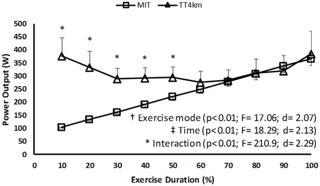
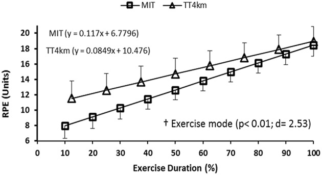
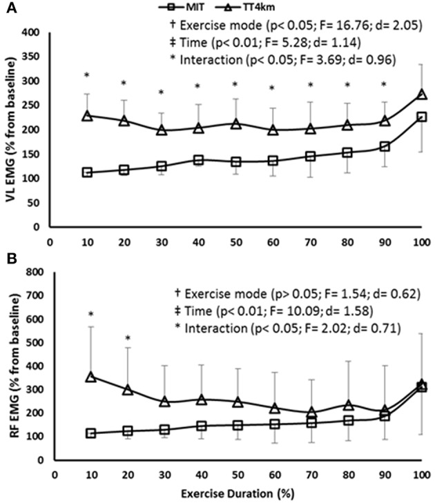
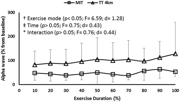
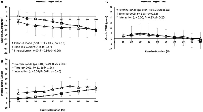
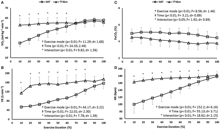
We investigated cerebral responses, simultaneously with peripheral and ratings of perceived exertion (RPE) responses, during different VO2MAX-matched aerobic exercise modes. Nine cyclists (VO2MAX of 57.5 ± 6.2 ml·kg(-1)·min(-1)) performed a maximal, controlled-pace incremental test (MIT) and a self-paced 4 km time trial (TT4km). Measures of cerebral (COX) and muscular (MOX) oxygenation were assessed throughout the exercises by changes in oxy- (O2Hb) and deoxy-hemoglobin (HHb) concentrations over the prefrontal cortex (PFC) and vastus lateralis (VL) muscle, respectively. Primary motor cortex (PMC) electroencephalography (EEG), VL, and rectus femoris EMG were also assessed throughout the trials, together with power output and cardiopulmonary responses. The RPE was obtained at regular intervals. Similar motor output (EMG and power output) occurred from 70% of the duration in MIT and TT4km, despite the greater motor output, muscle deoxygenation (↓ MOX) and cardiopulmonary responses in TT4km before that point. Regarding cerebral responses, there was a lower COX (↓ O2Hb concentrations in PFC) at 20, 30, 40, 50 and 60%, but greater at 100% of the TT4km duration when compared to MIT. The alpha wave EEG in PMC remained constant throughout the exercise modes, with greater values in TT4km. The RPE was maximal at the endpoint in both exercises, but it increased slower in TT4km than in MIT. Results showed that similar motor output and effort tolerance were attained at the closing stages of different VO2MAX-matched aerobic exercises, although the different disturbance until that point. Regardless of different COX responses during most of the exercises duration, activation in PMC was preserved throughout the exercises, suggesting that these responses may be part of a centrally-coordinated exercise regulation.
我们在不同的与最大摄氧量(VO2MAX)匹配的有氧运动模式下,同时研究了大脑反应以及外周反应和自觉用力程度(RPE)反应。九名自行车运动员(VO2MAX为57.5±6.2毫升·千克⁻¹·分钟⁻¹)进行了一次最大强度、控制节奏的递增测试(MIT)和一次自定节奏的4公里计时赛(TT4km)。在整个运动过程中,分别通过前额叶皮层(PFC)上氧合血红蛋白(O2Hb)和脱氧血红蛋白(HHb)浓度的变化来评估大脑(COX)和肌肉(MOX)的氧合情况。在整个试验过程中还评估了初级运动皮层(PMC)脑电图(EEG)、股外侧肌(VL)和股直肌肌电图,以及功率输出和心肺反应。定期获取RPE。尽管在TT4km中该点之前运动输出、肌肉脱氧(↓MOX)和心肺反应更大,但在MIT和TT4km中从70%的持续时间起出现了相似的运动输出(肌电图和功率输出)。关于大脑反应,与MIT相比,在TT4km的20%、30%、40%、50%和60%时COX较低(PFC中O2Hb浓度↓),但在100%的TT4km持续时间时较高。在整个运动模式中,PMC中的α波脑电图保持恒定,在TT4km中值更大。在两种运动中,RPE在终点时最大,但在TT4km中比在MIT中增加得慢。结果表明,在不同的与VO2MAX匹配的有氧运动的结束阶段,尽管在此之前有不同的干扰,但仍能获得相似的运动输出和耐力。在大多数运动持续时间内,尽管COX反应不同,但在整个运动过程中PMC的激活得以保留,这表明这些反应可能是中枢协调运动调节的一部分。